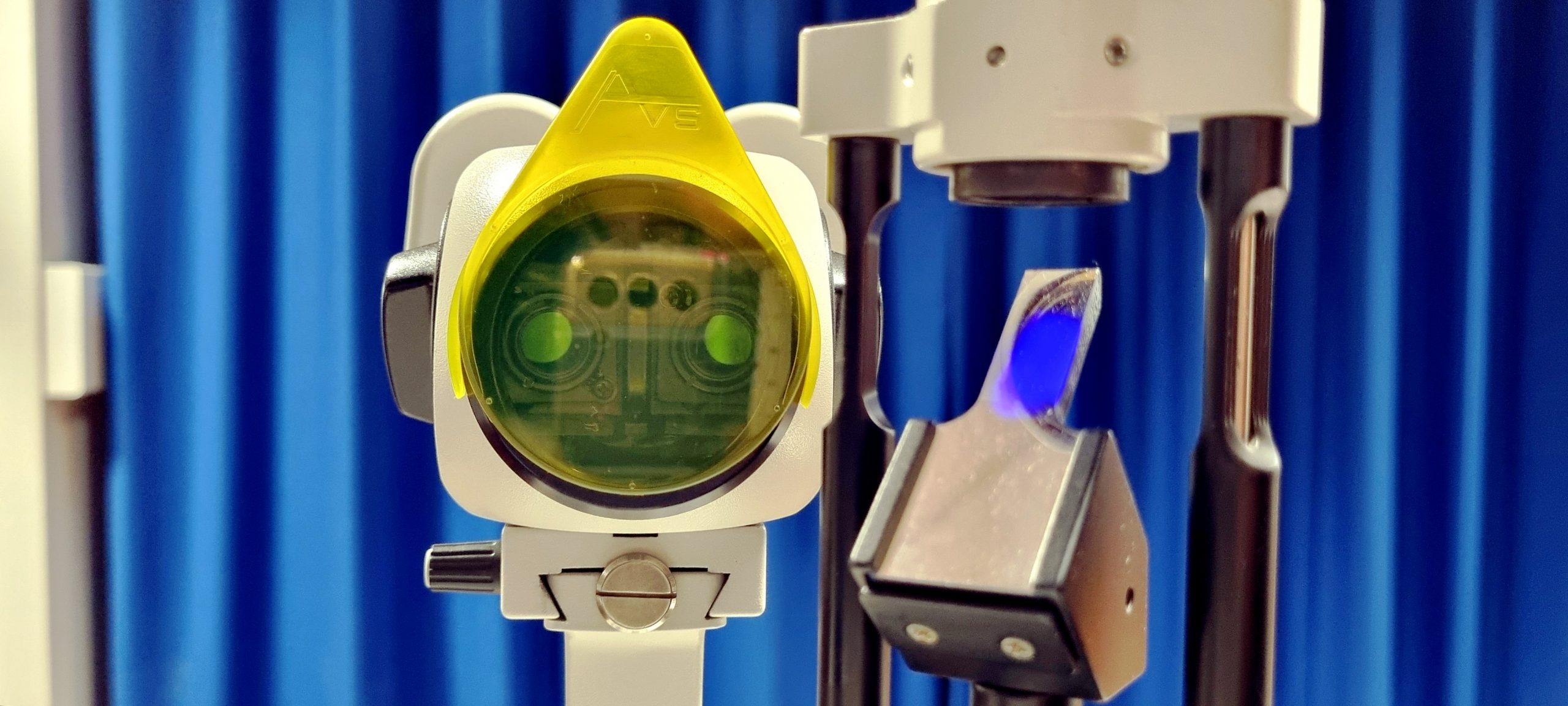
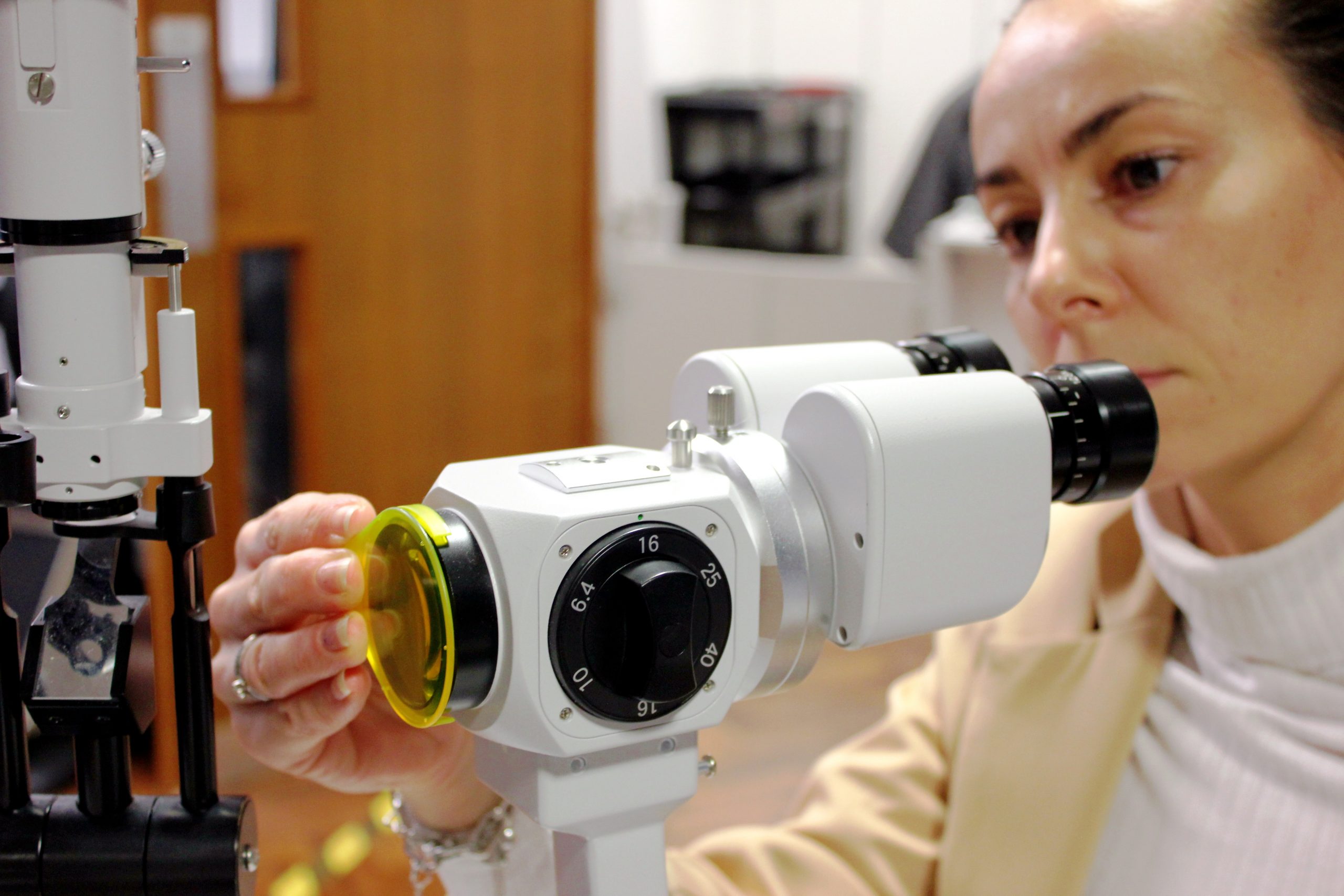
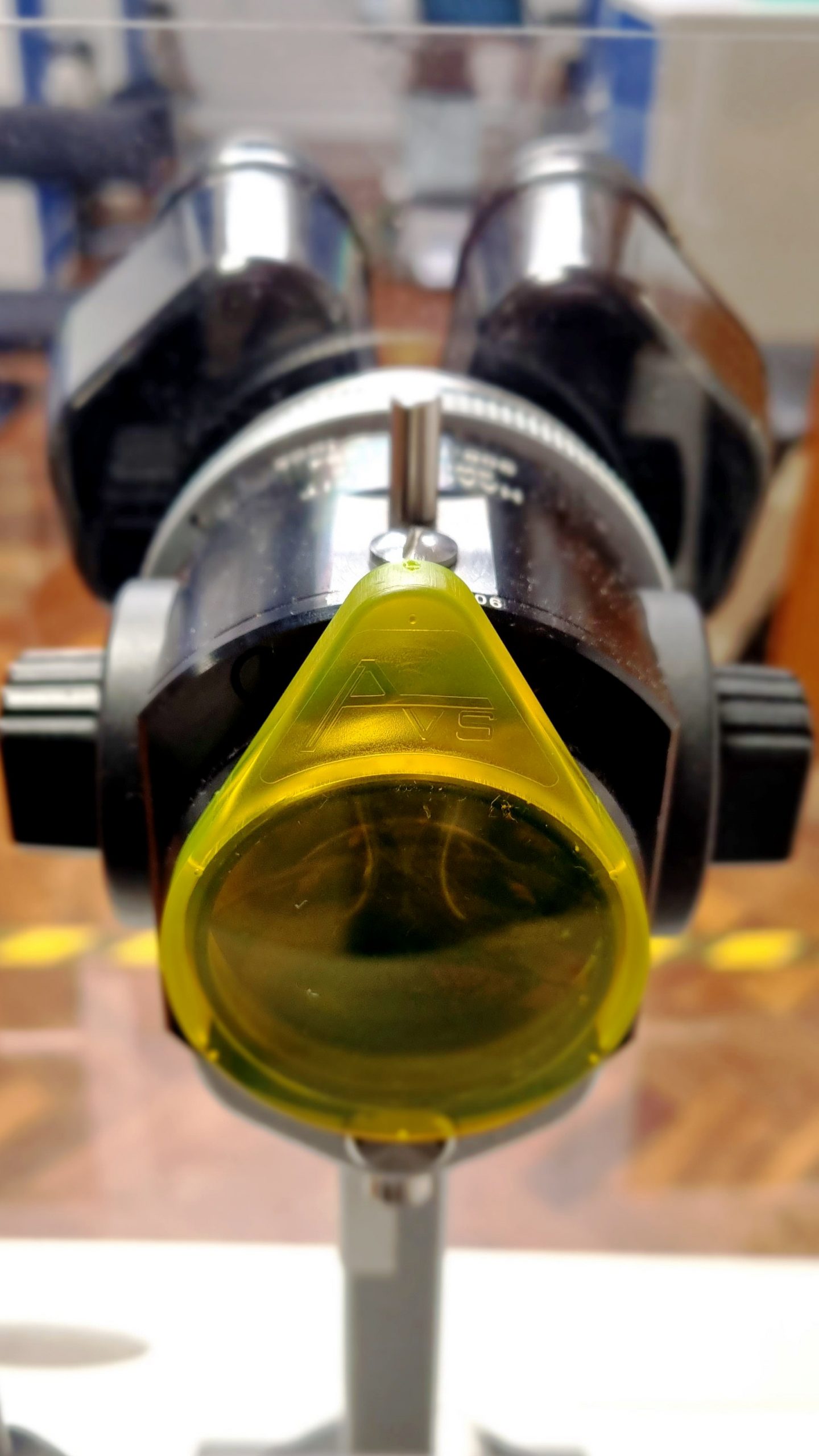
Front-on view of the Aston Vision Sciences fluorescein filter mounted on a CSO slit lamp, highlighting its use in detailed eye examinations
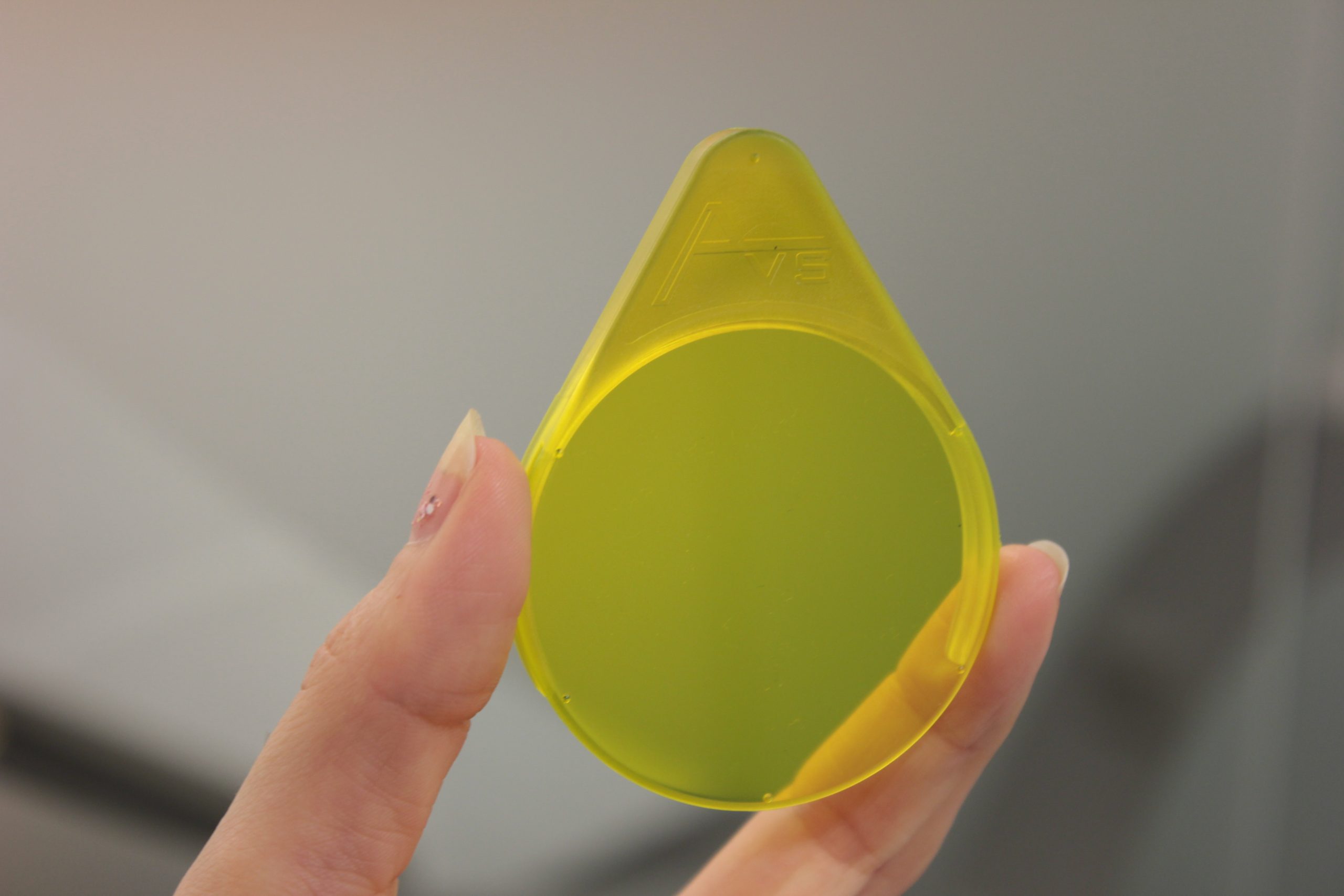
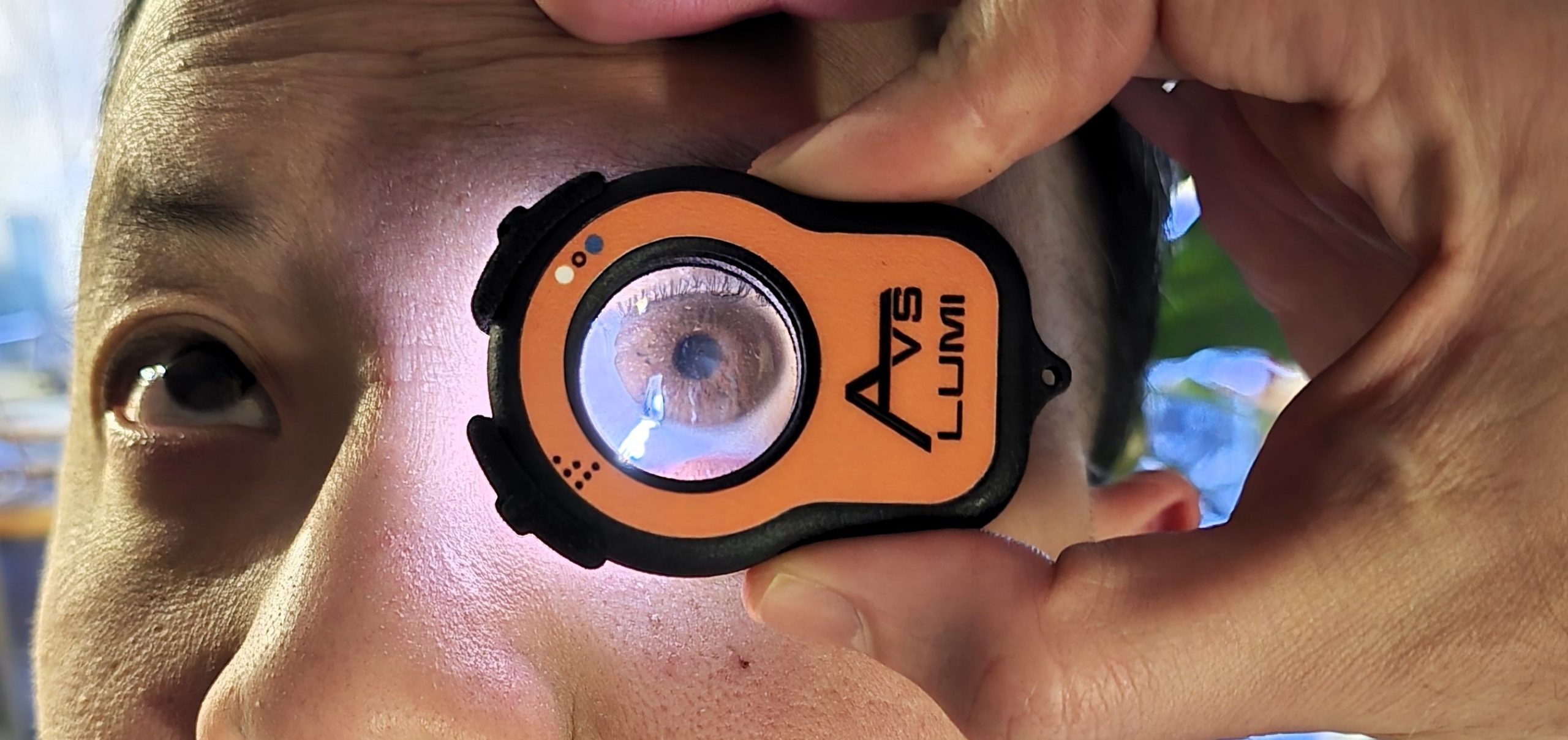
A user holding the AVS fluorescein filter
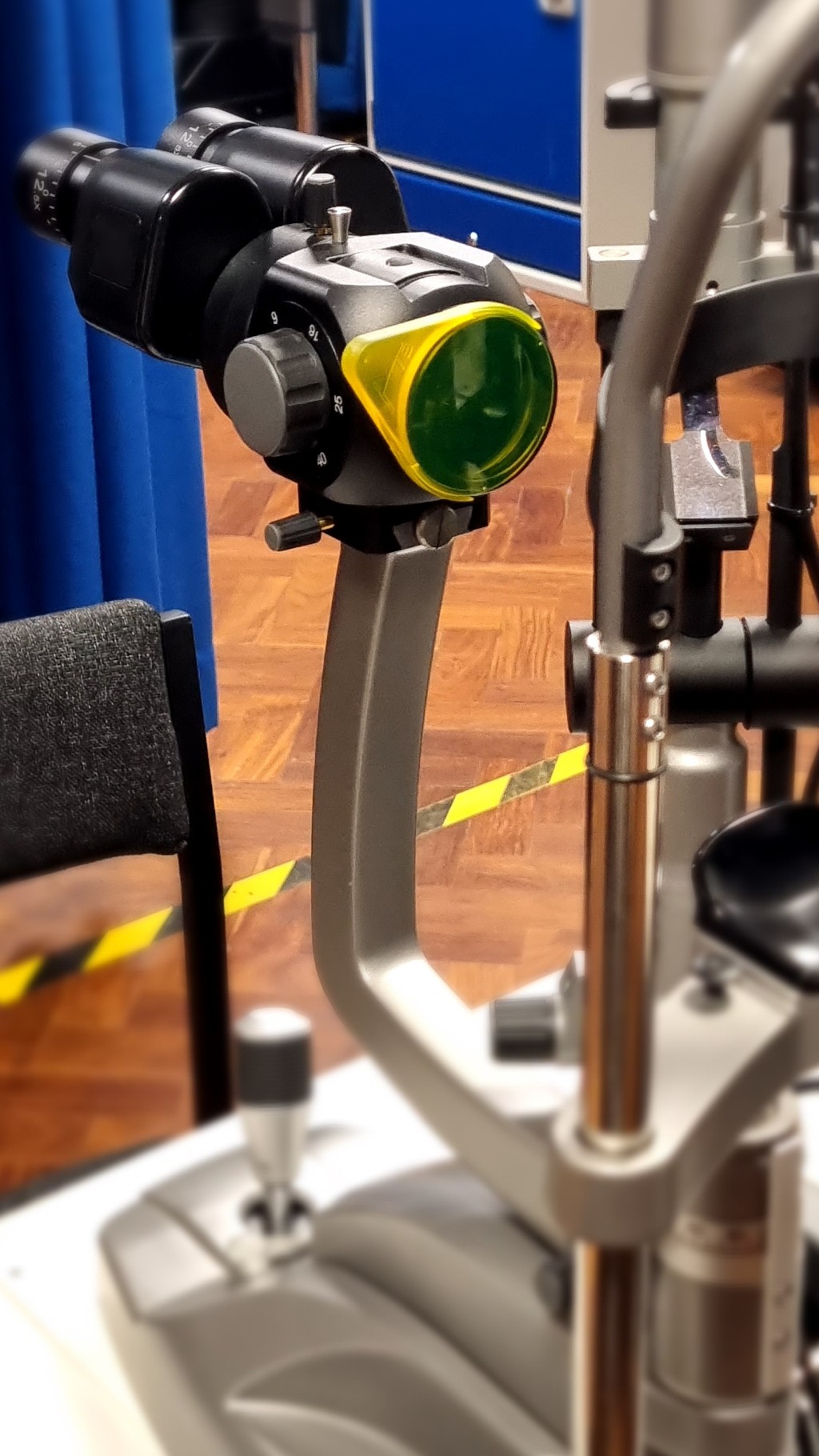
AVS Watten filter that can be used for anterior and dry eye evaluations
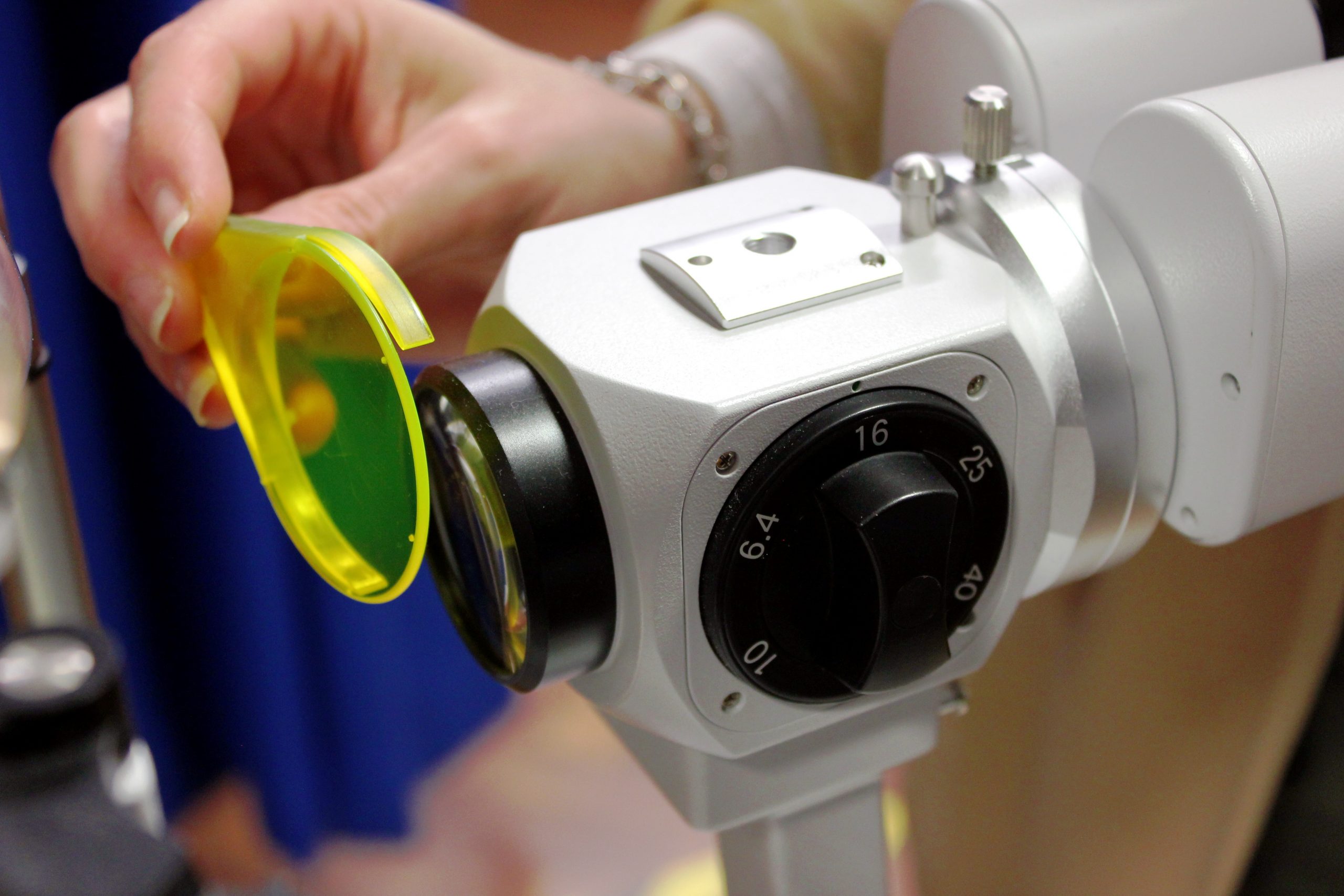
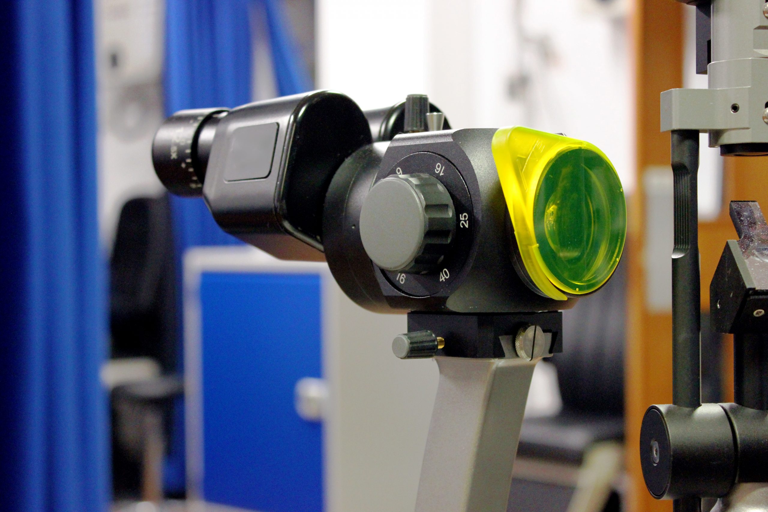
Fluorescein filter about to be clipped on to the slit lamp objective lens fo hands-fee use
Filter can be held in front of the slit lamp objective lens to enable Fluorescein observation on non compatible models.
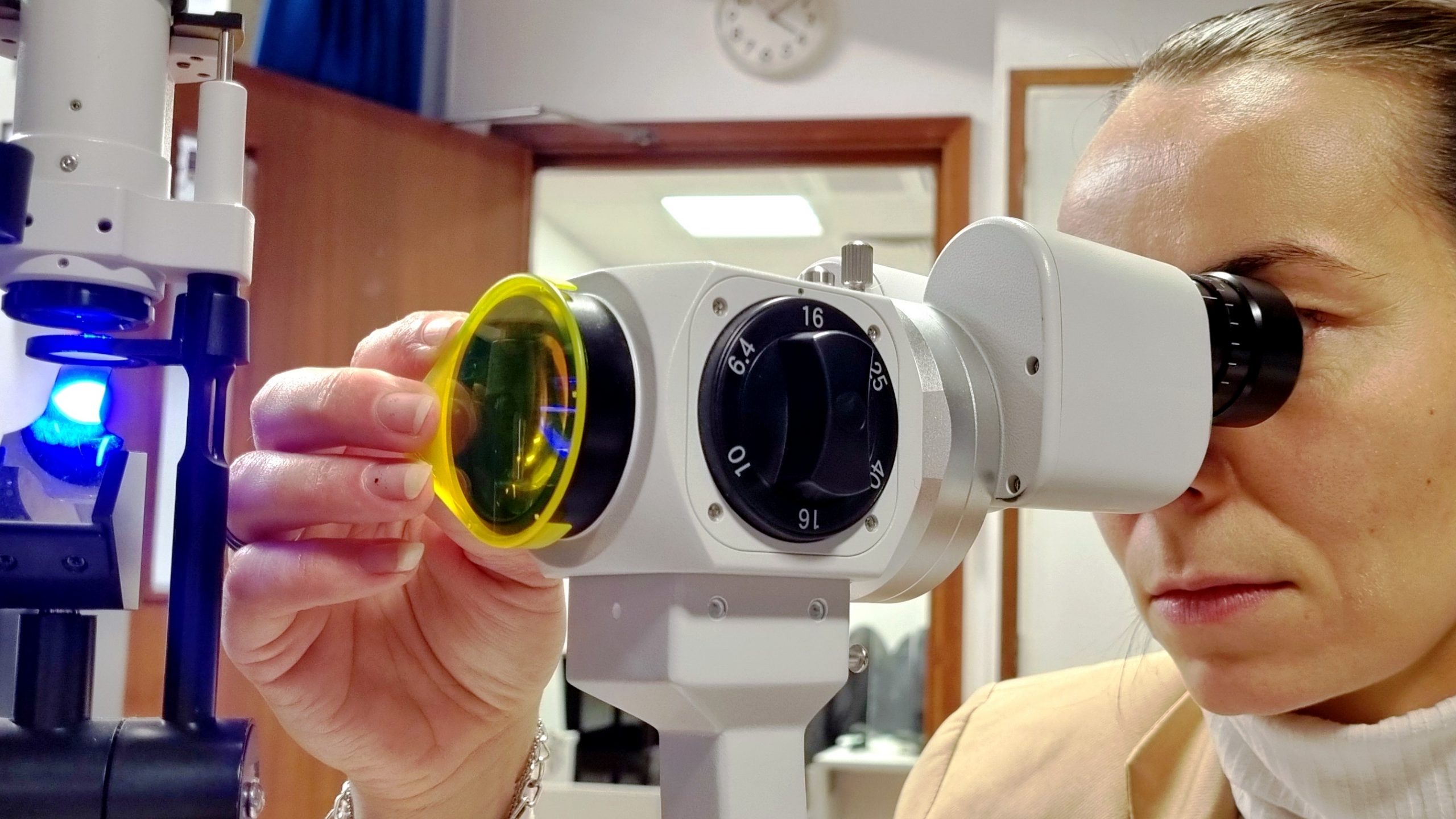
Fluorescein Filter being attached to slit lamp
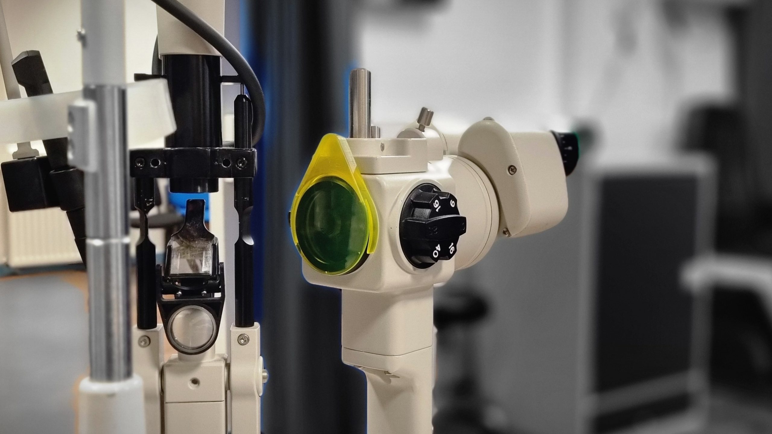
Fluorescein Filter on Keeler 40H Slit Lamp
The AVS fluorescein filter attached to a Keeler slit lamp enables improved visualization of fluorescein dye during anterior eye examinations
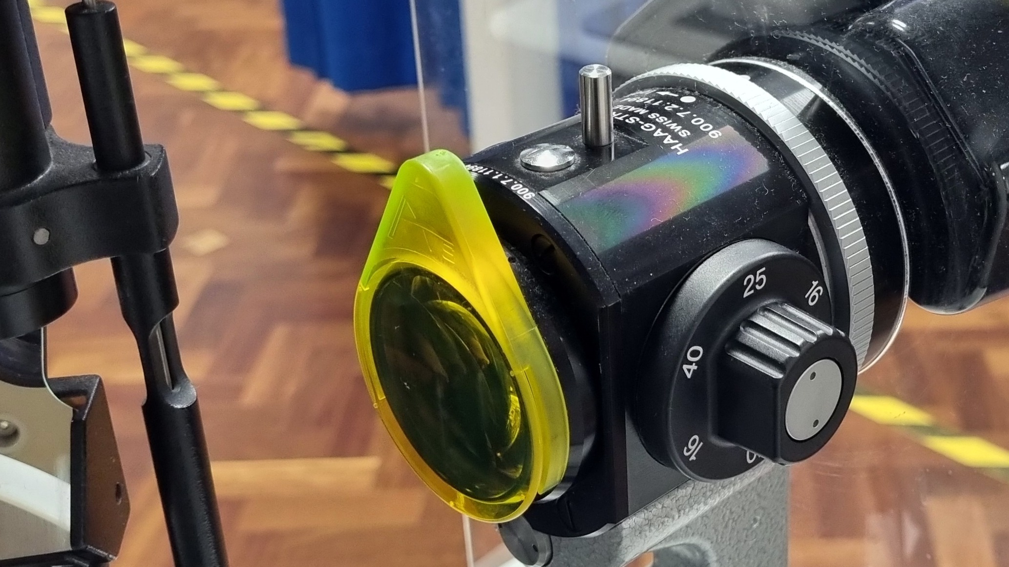
AVS Fluorescein Filter attaced to a Haag Streit slit lamp
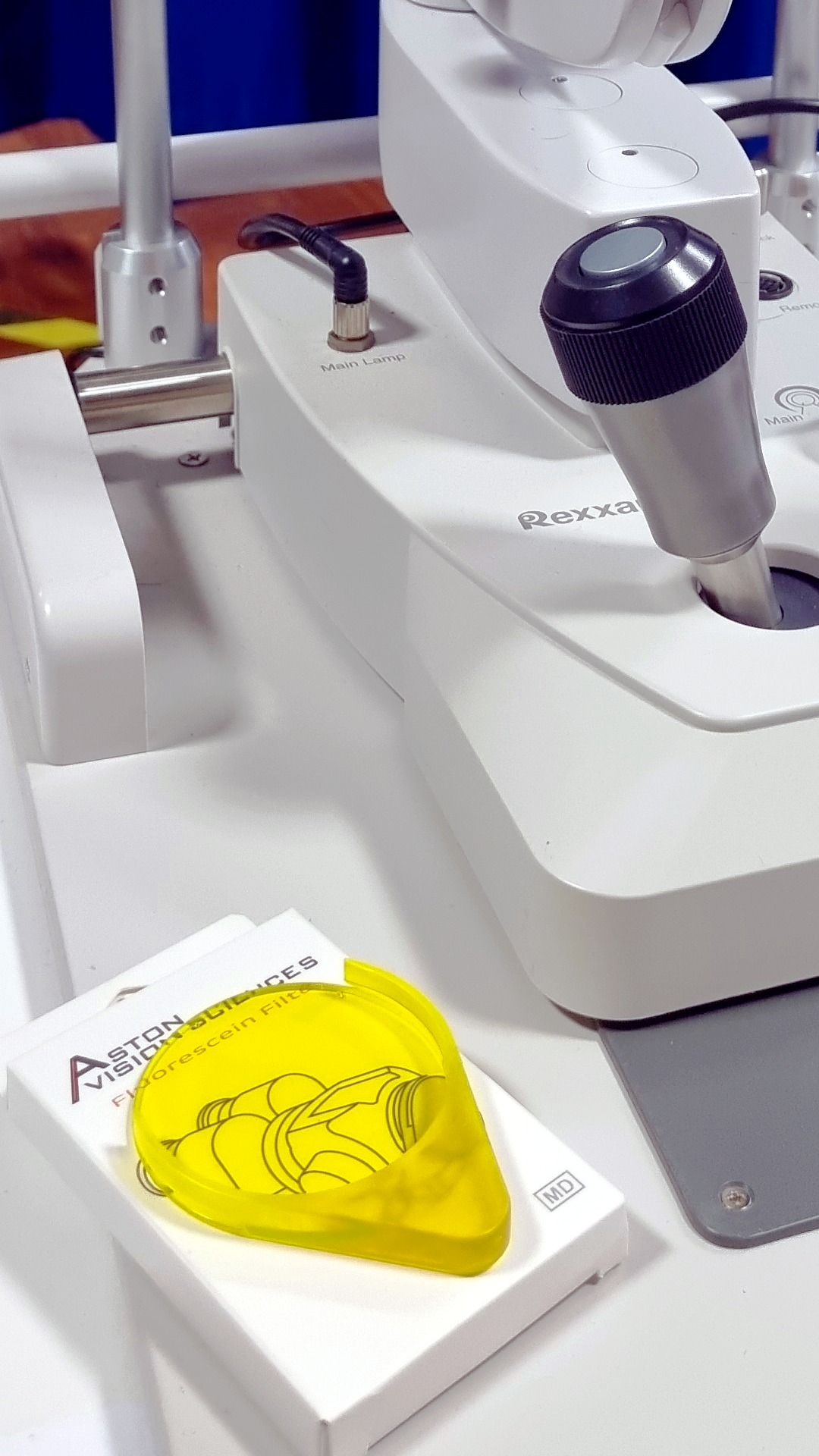
Fluorescein Filter is an optometry tool that can fit on many slit lamps
Gold standard Fluorescein viewing from Aston Vision Sciences
Fluorescein filter ready to be used for foreign body detection
Wratten filter to be used for dry eye, anterior eye and corneal evaluation.
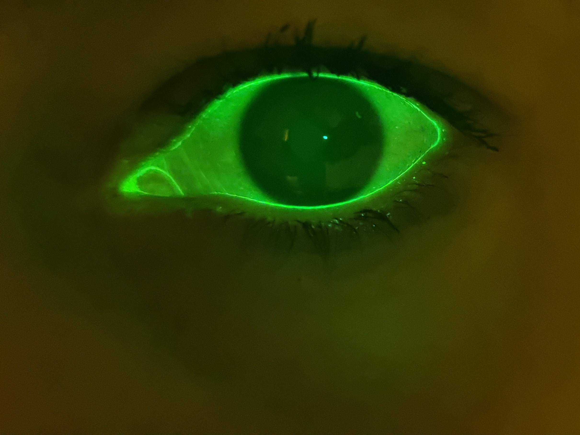
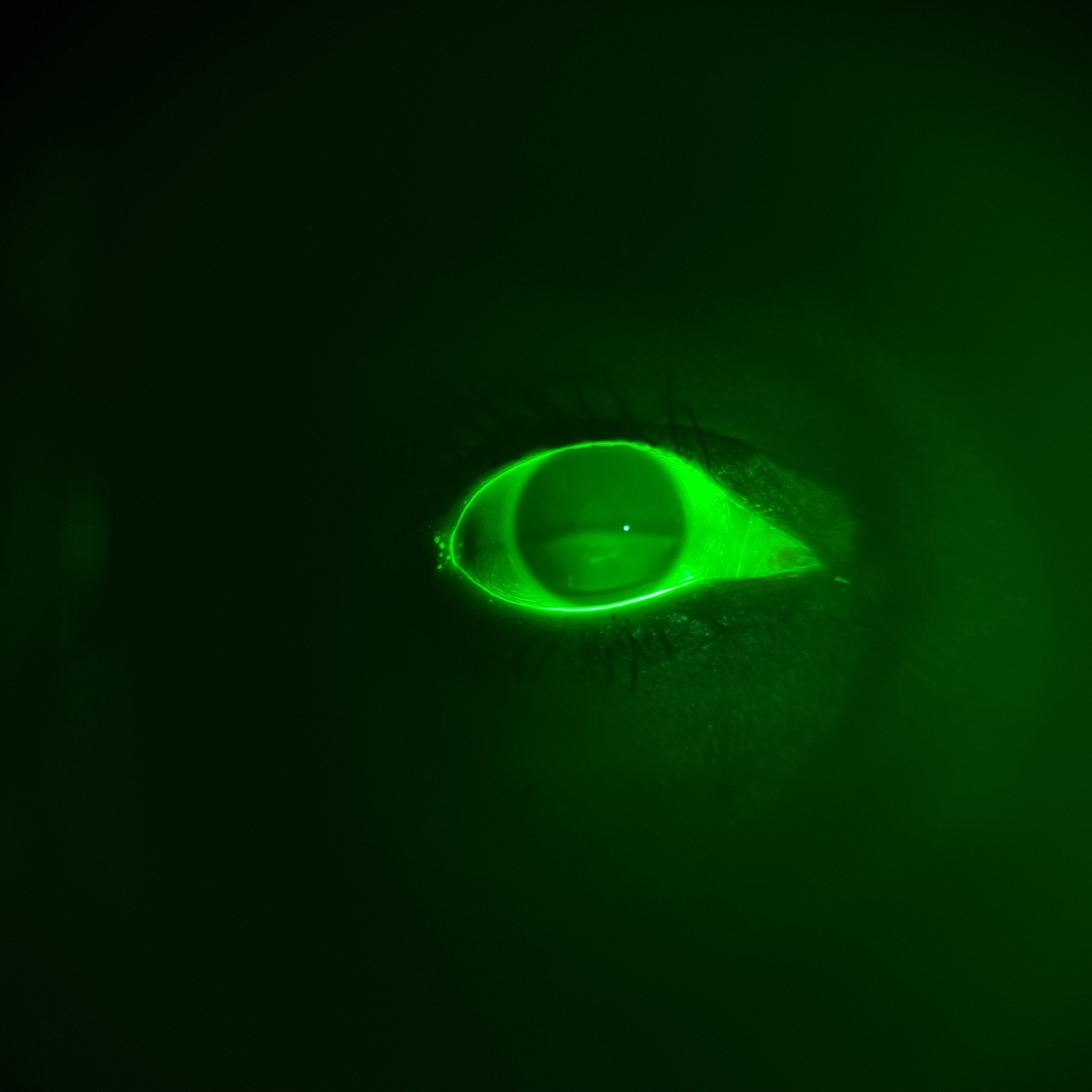
Close-up view of a human eye under green fluorescein illumination as viewed through the AVS Fluorescein Filter
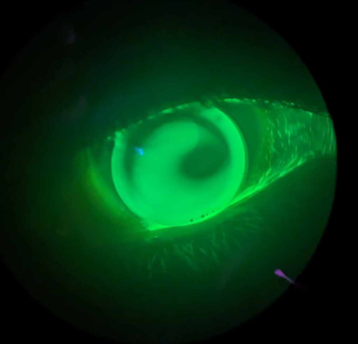
Tear film breakup in action.
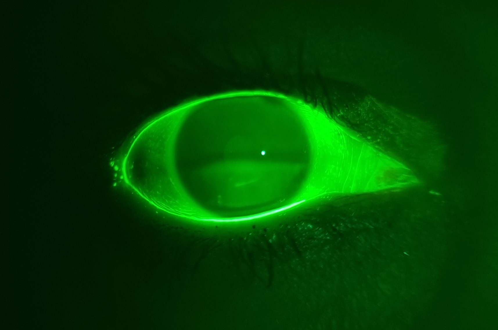
Scratch on inferior cornea
An eye illuminated with fluorescein dye under blue light reveals tear film instability, often caused by partial blinking — a key factor in dry eye disease
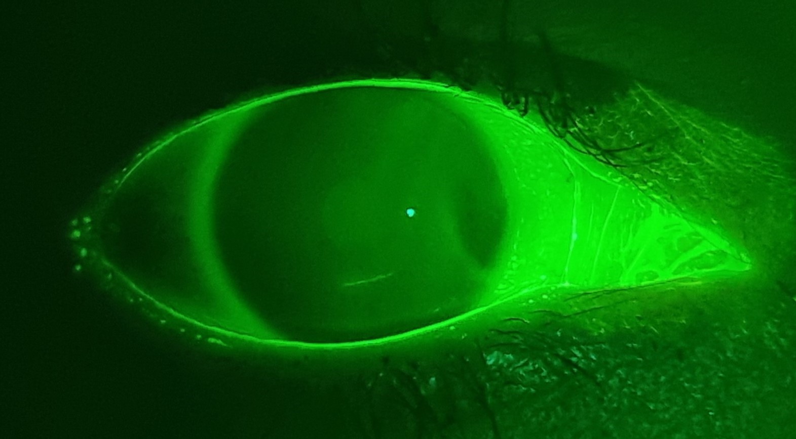
Fluorescein dye highlights the tear film and contact lens fit during a slit lamp examination, aiding in dry eye and anterior segment diagnostics
Aston Vision Sciences Fluorescein filter used to observe tear breakup in action giving an indication of tear stability over time
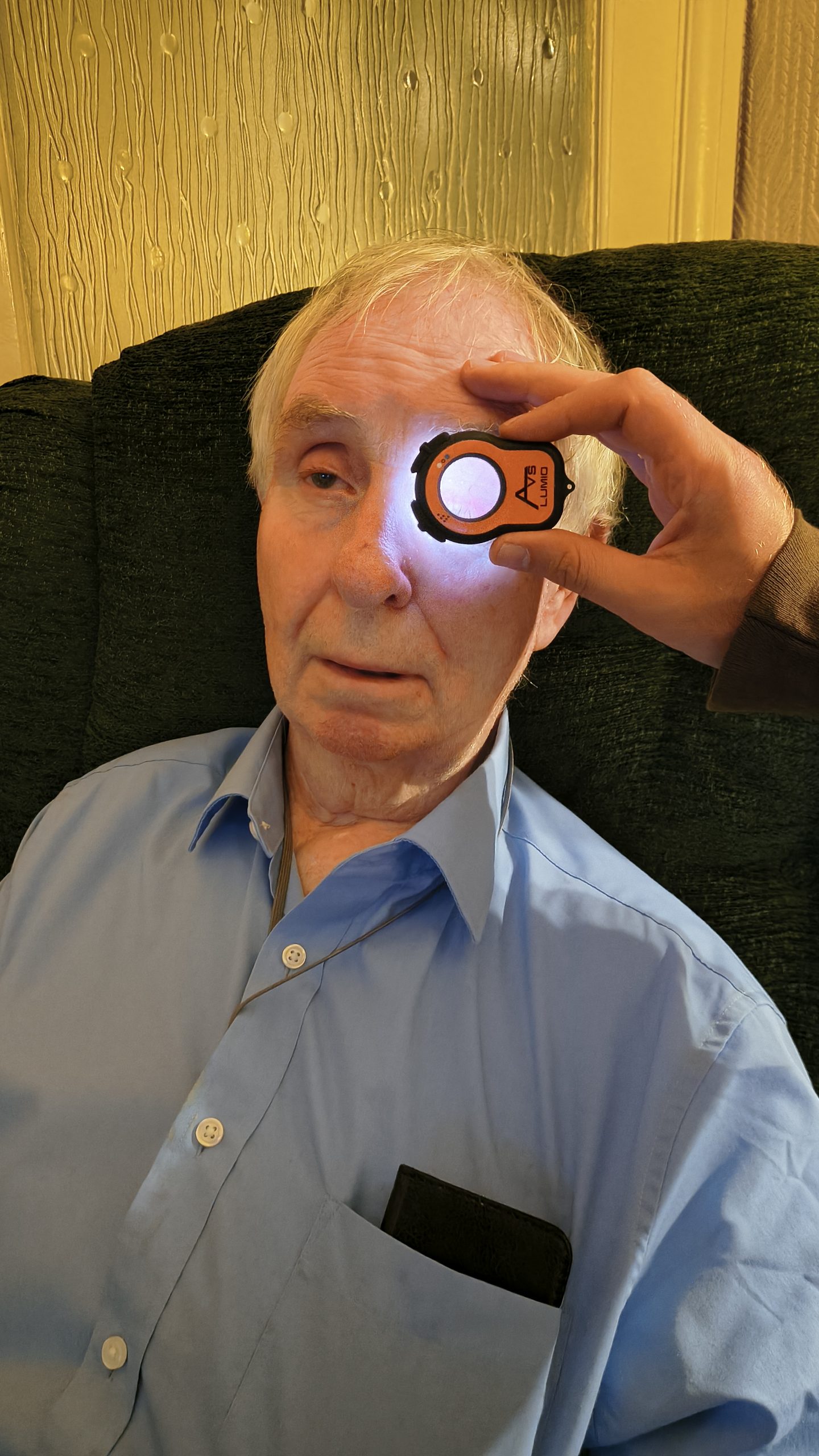
Speed and convenience allow for comfortable domiciliary eye examinations
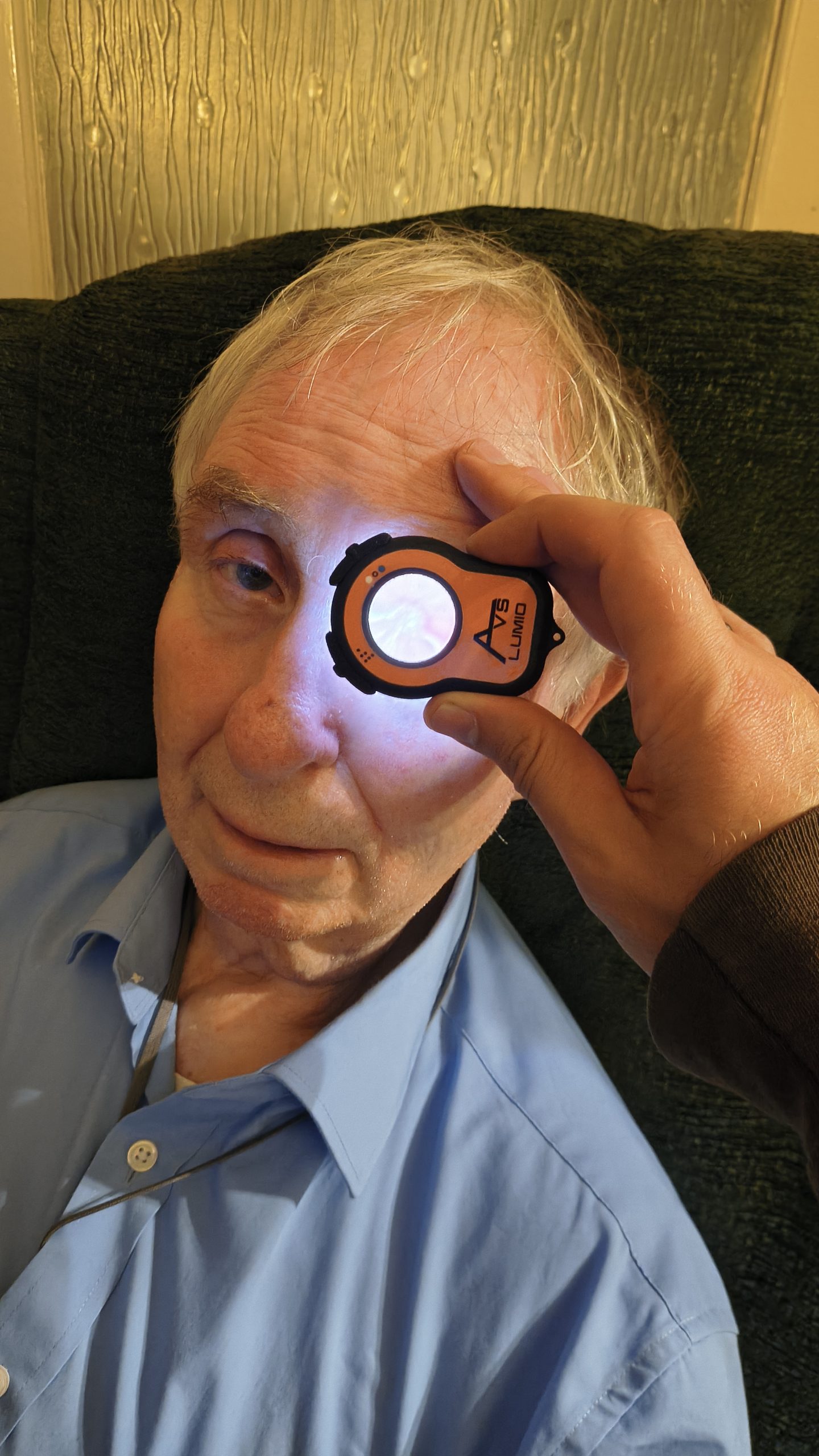
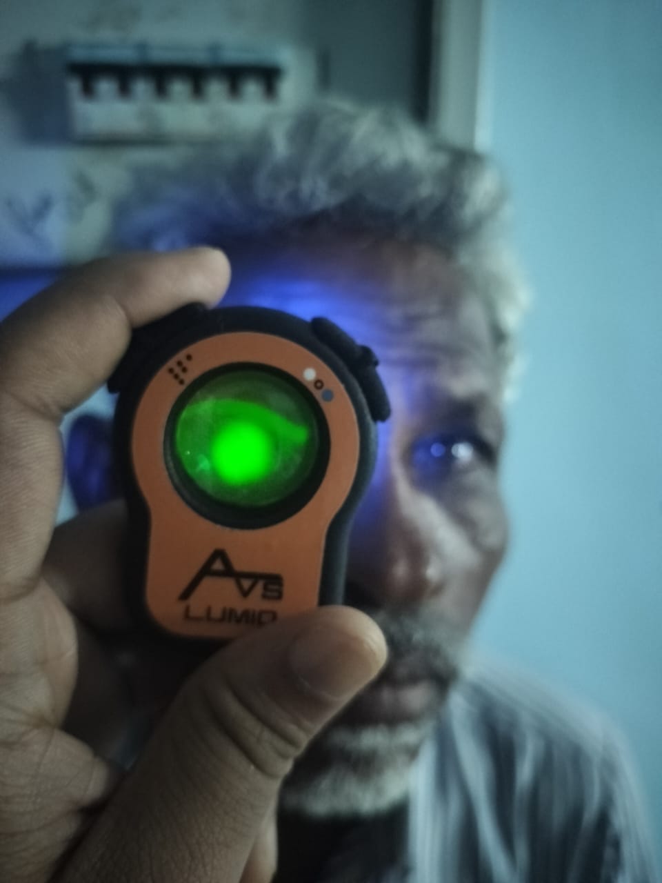
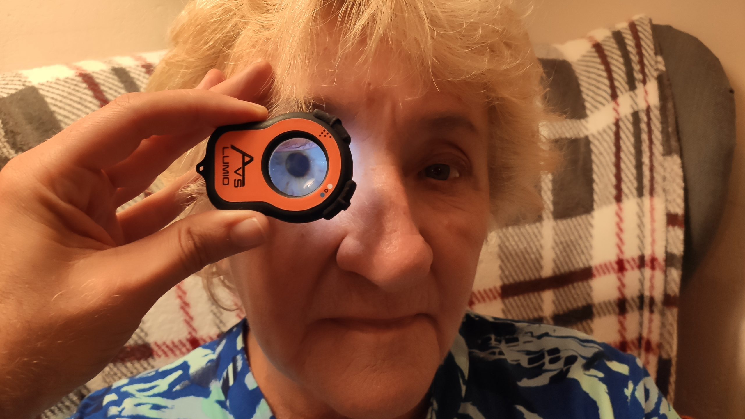
A close-up of an elderly patient undergoing a home-based eye exam using the Lumio device for anterior segment assessment.
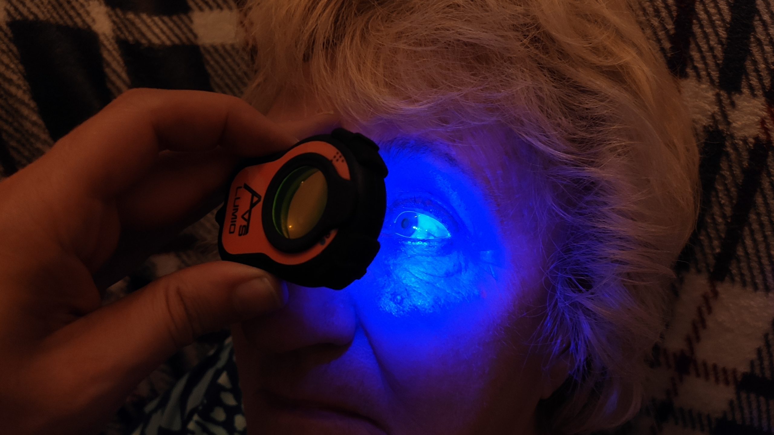
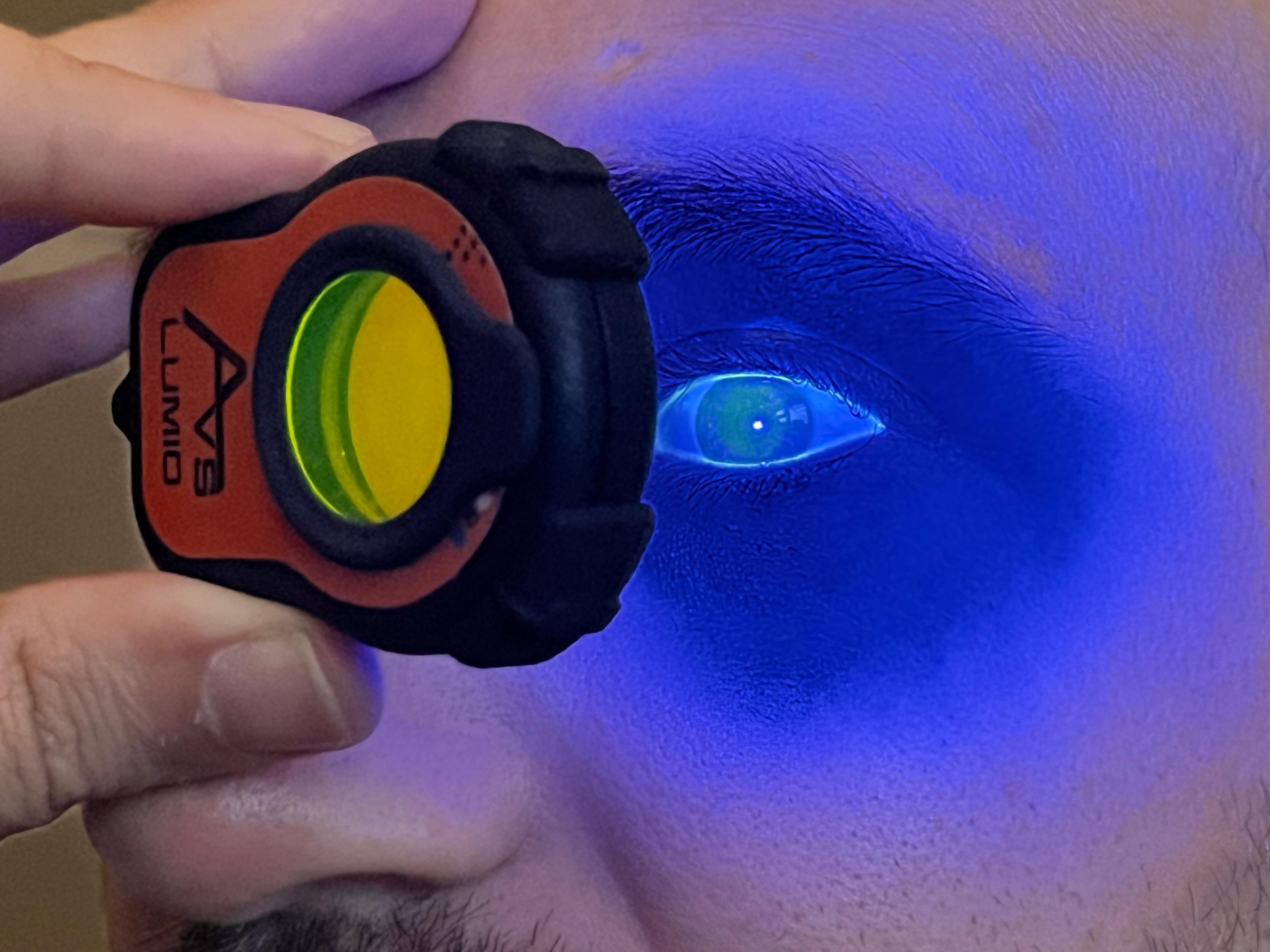
The Lumio device in action, emitting blue light in combination with the Fluorescein filter to examine the eye during a home visit examination.
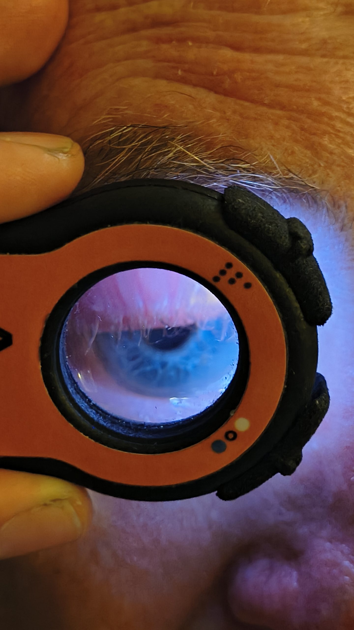
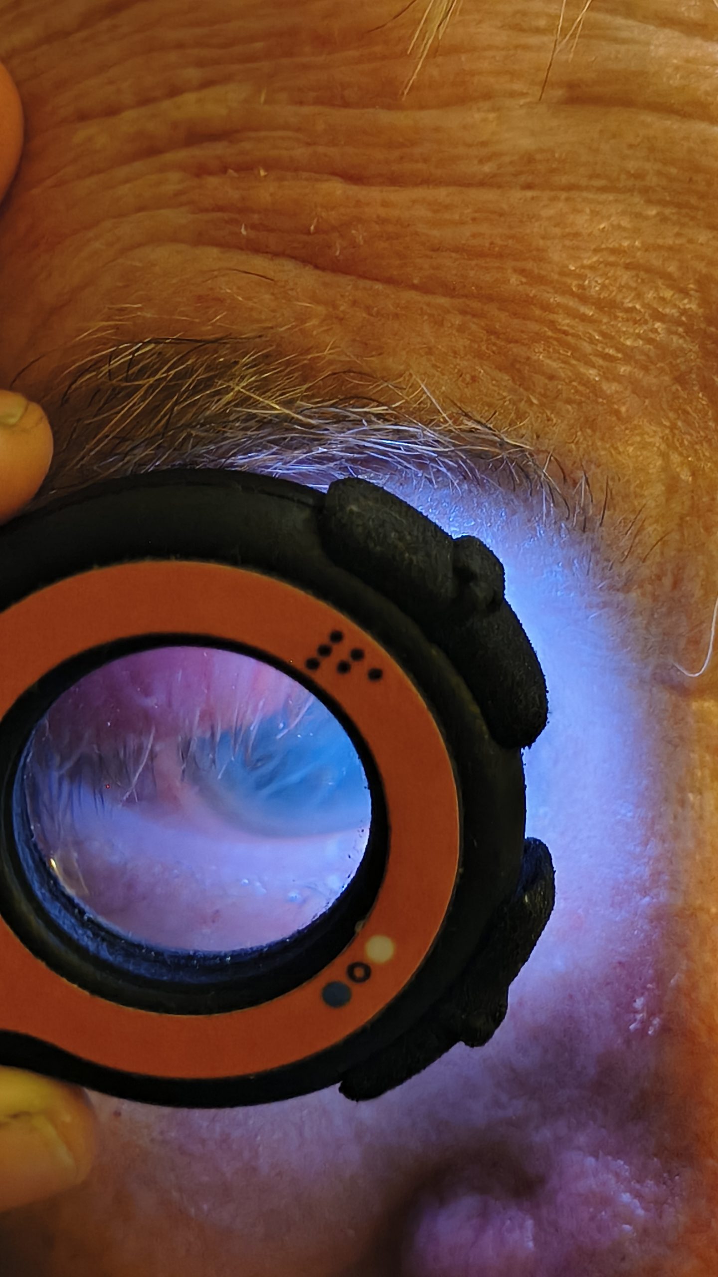
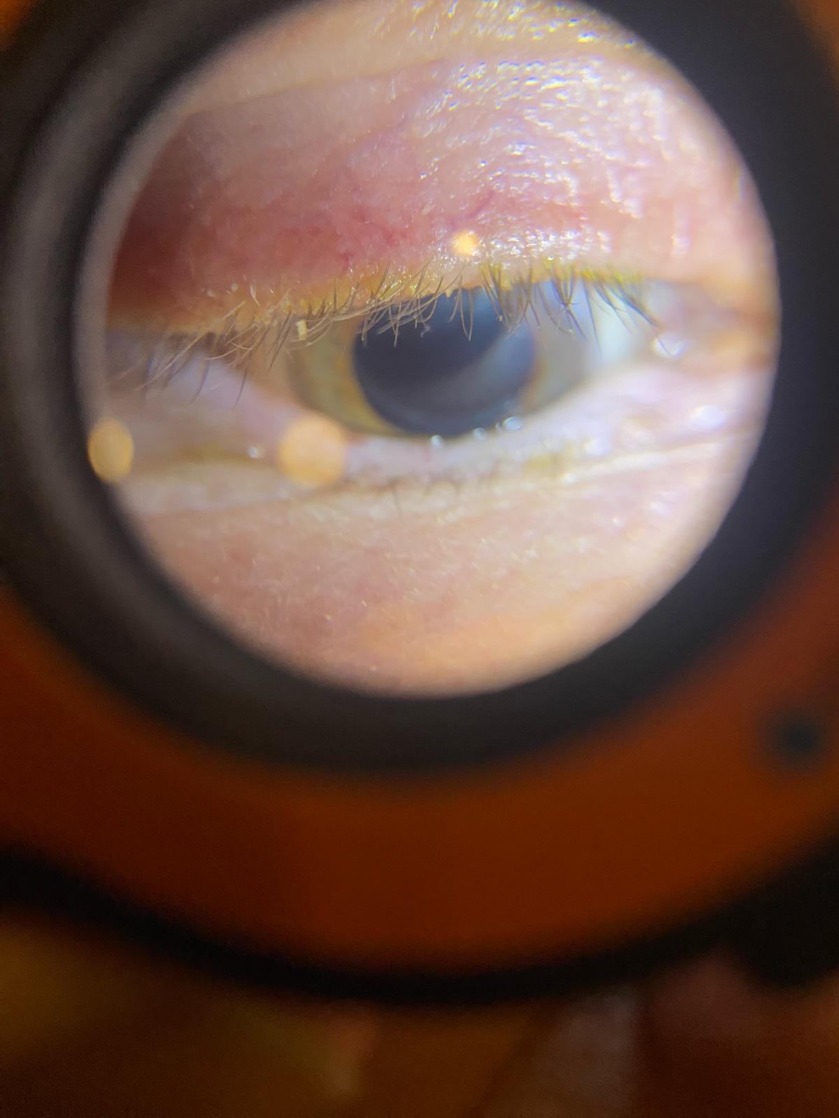
A close-up view through the Lumio device lens reveals detailed features of the patient’s eye during a home exam.
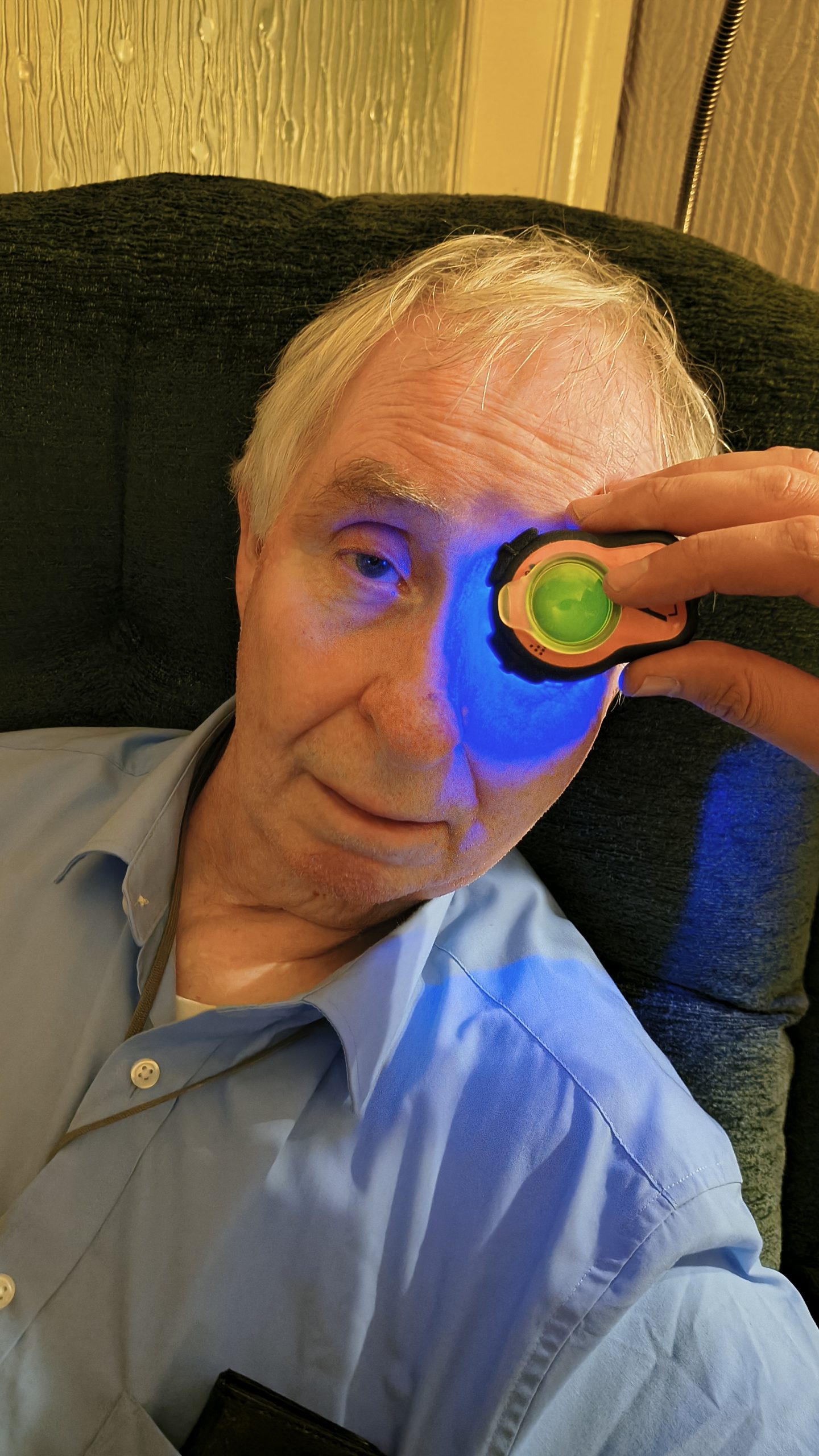
A focused view of an elderly patient’s eye captured through the Lumio device during a domiciliary optometry session.
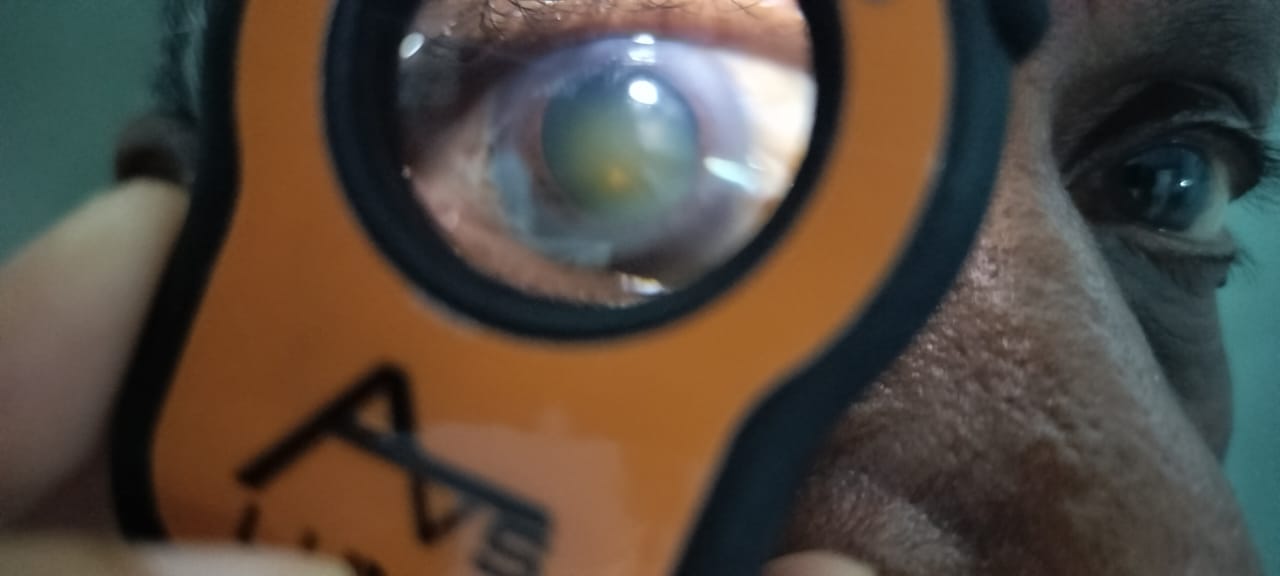
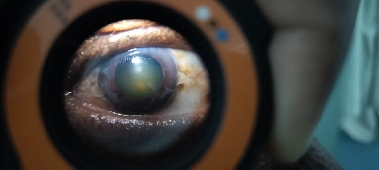
A cataract-affected eye examined using Lumio, showing lens opacity and structural changes
A dilated pupil reveals cataract changes in the lens, visualized with Lumio’s diagnostic clarity
Accessible Eye Care for All: Advanced Diagnostics with Lumio
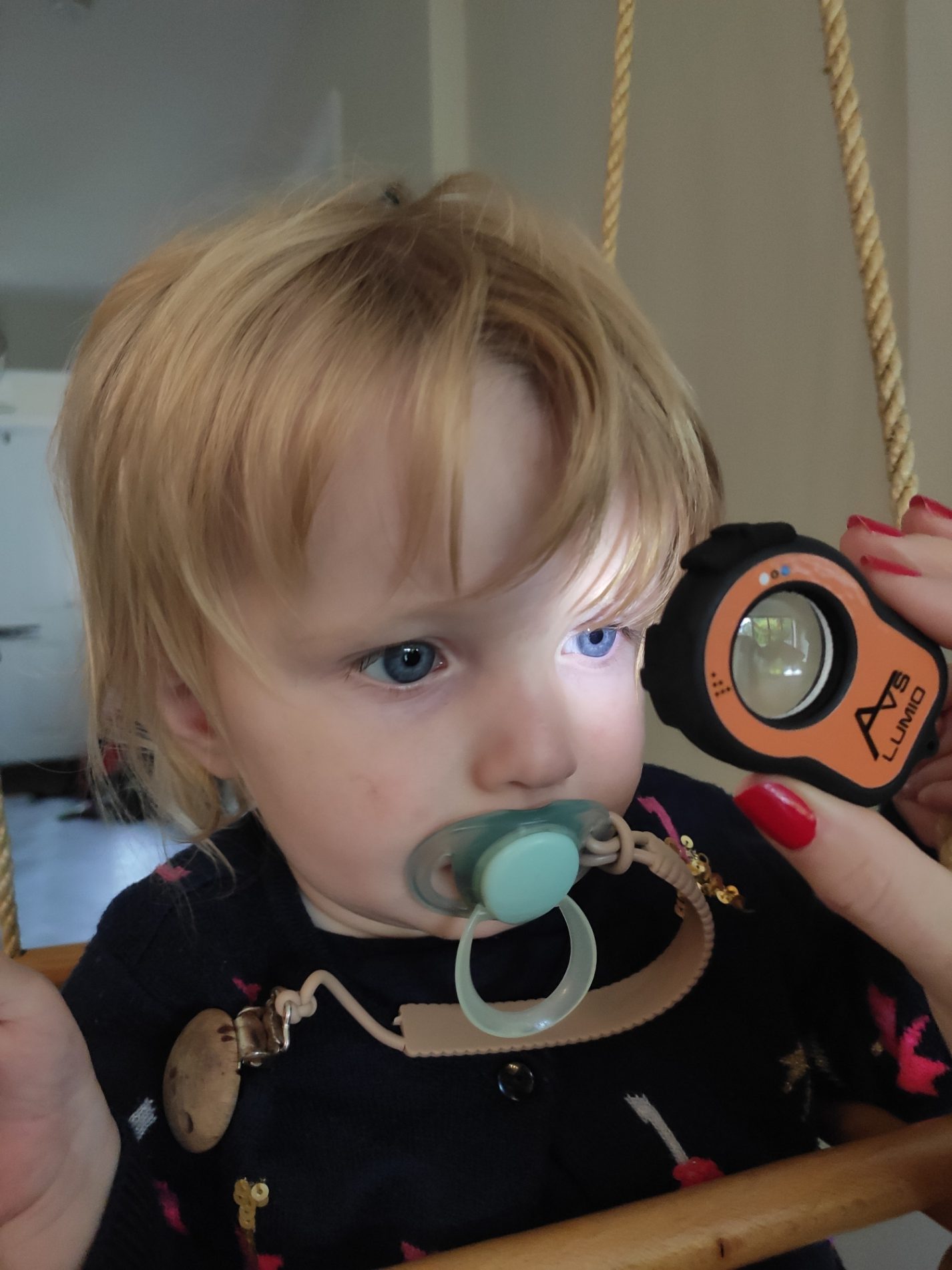
Pediatric Eye Exam with Lumio Fluorescein – Advanced Diagnostics for Young Eyes
A young child engages with the Lumio diagnostic device during a home visit, highlighting Aston Vision’s family-friendly approach to eye care
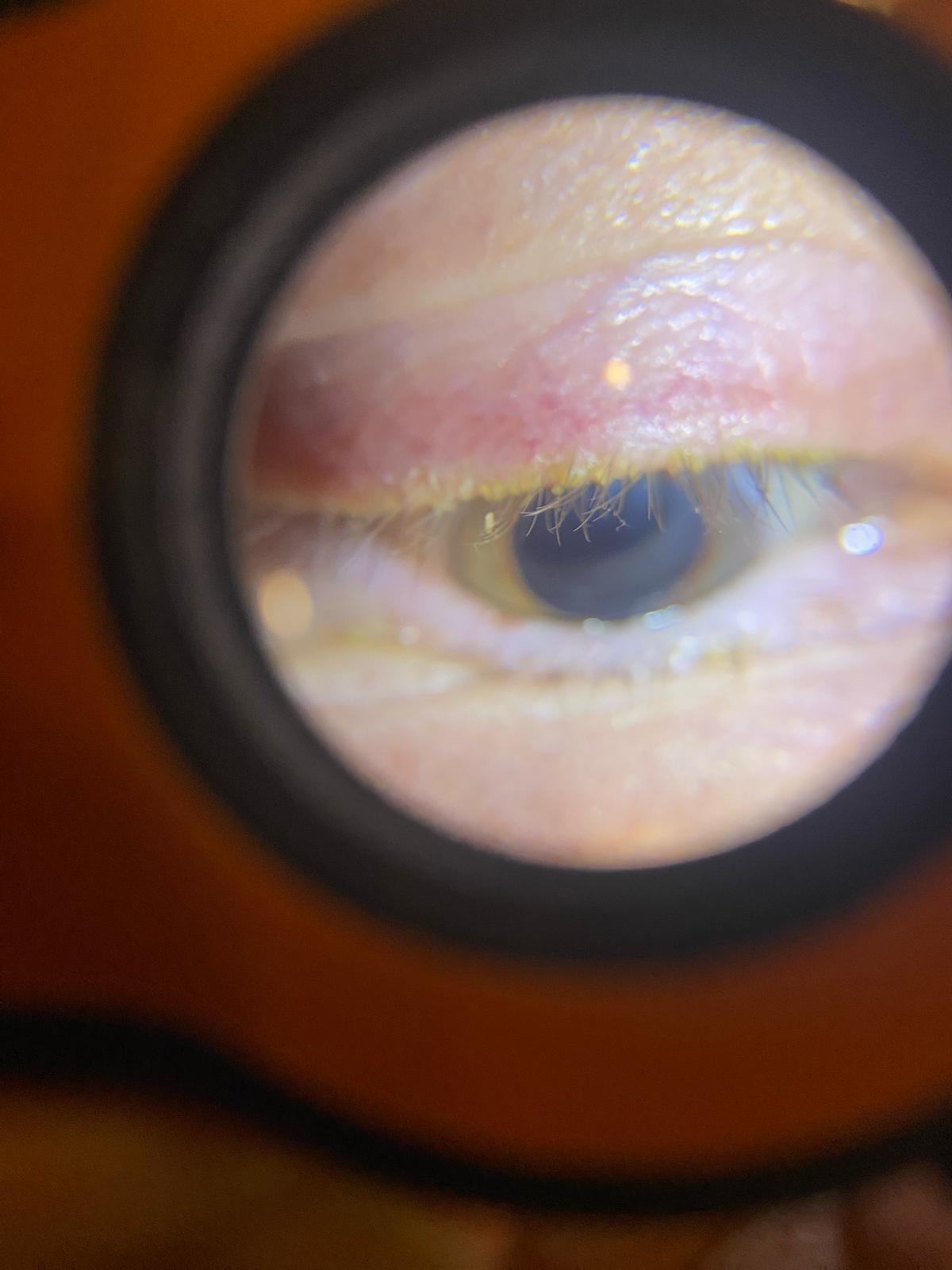
A close-up of an eye being examined with the Lumio device, demonstrating its precision in anterior segment diagnostics.
A detailed view of an eyelid affected by Demodex blepharitis, captured using the Lumio diagnostic device.
Another close-up of Demodex blepharitis, showing lash base inflammation and debris under Lumio examination.
Fluorescein viewing on a patient with suspected Tachoma
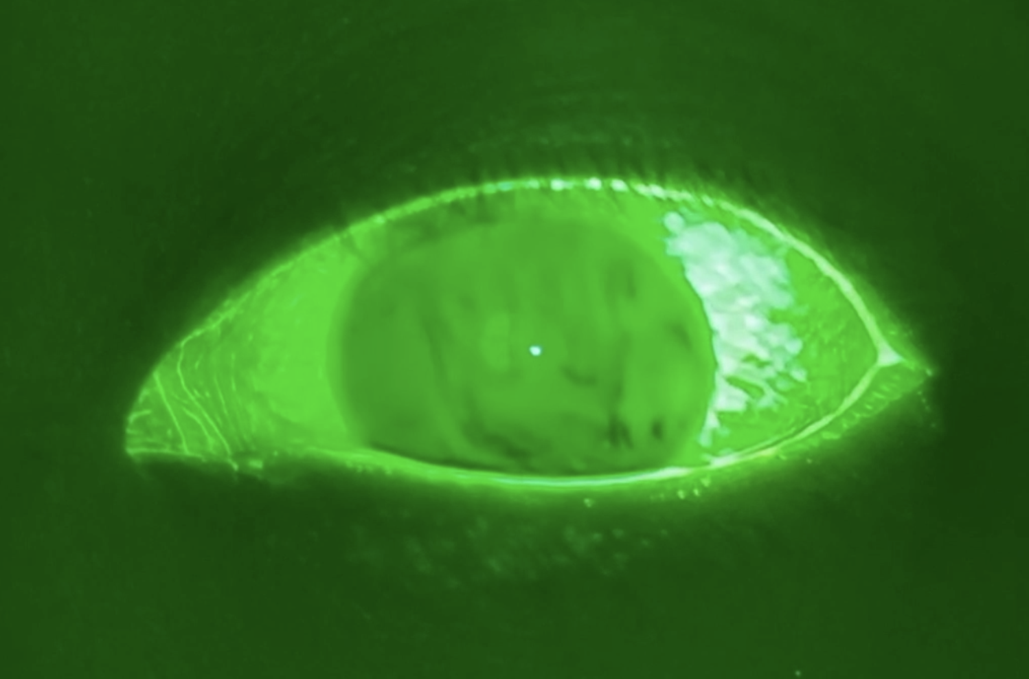
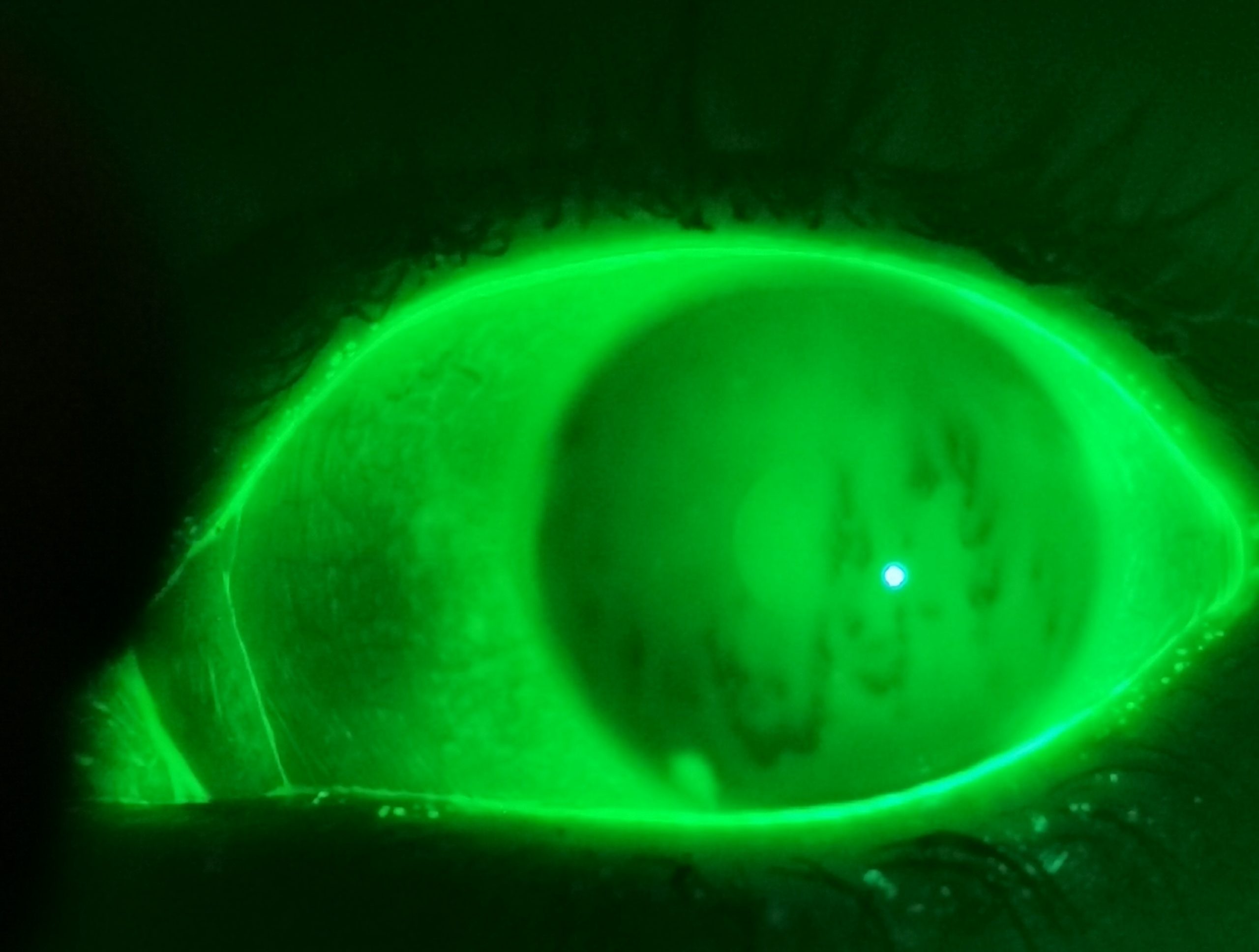
Fluorescein dye highlights punctate staining on the cornea, highlighting possible presence of corneal damage due to dry eye disesase.
Lumio used to examine patient eyelids and possible presence of ingrowing eyelashes
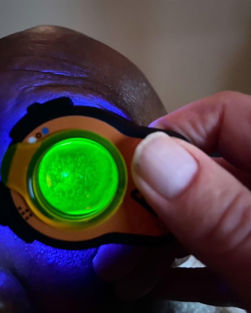
A close-up view of the Lumio device emitting blue light during a home eye exam, enhancing visibility of the ocular surface during Fluorescein examination
Advanced Eye Examination with Lumio & Fluorescein – Enhancing Diagnostics in Ophthalmology
Lumio device used in a home setting to illuminate the eye with blue light, revealing detailed ocular structures for fluorescein dry eye assessment
Pediatric Eye Examination with Lumio Under White Light – Safe and Precise Diagnostics for Infants
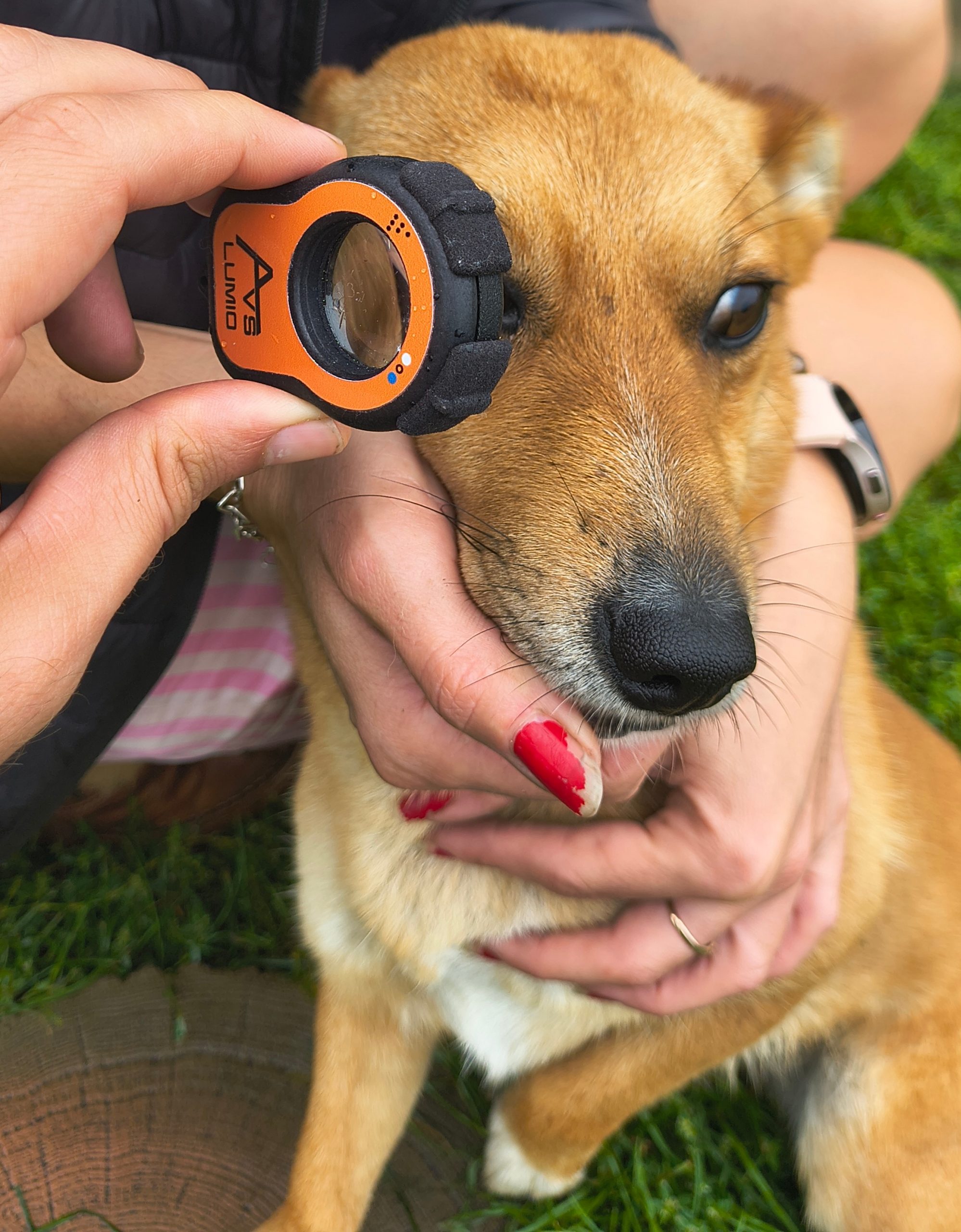
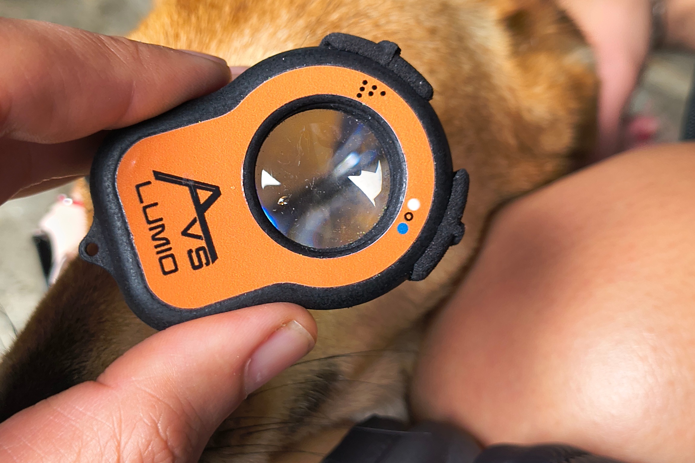
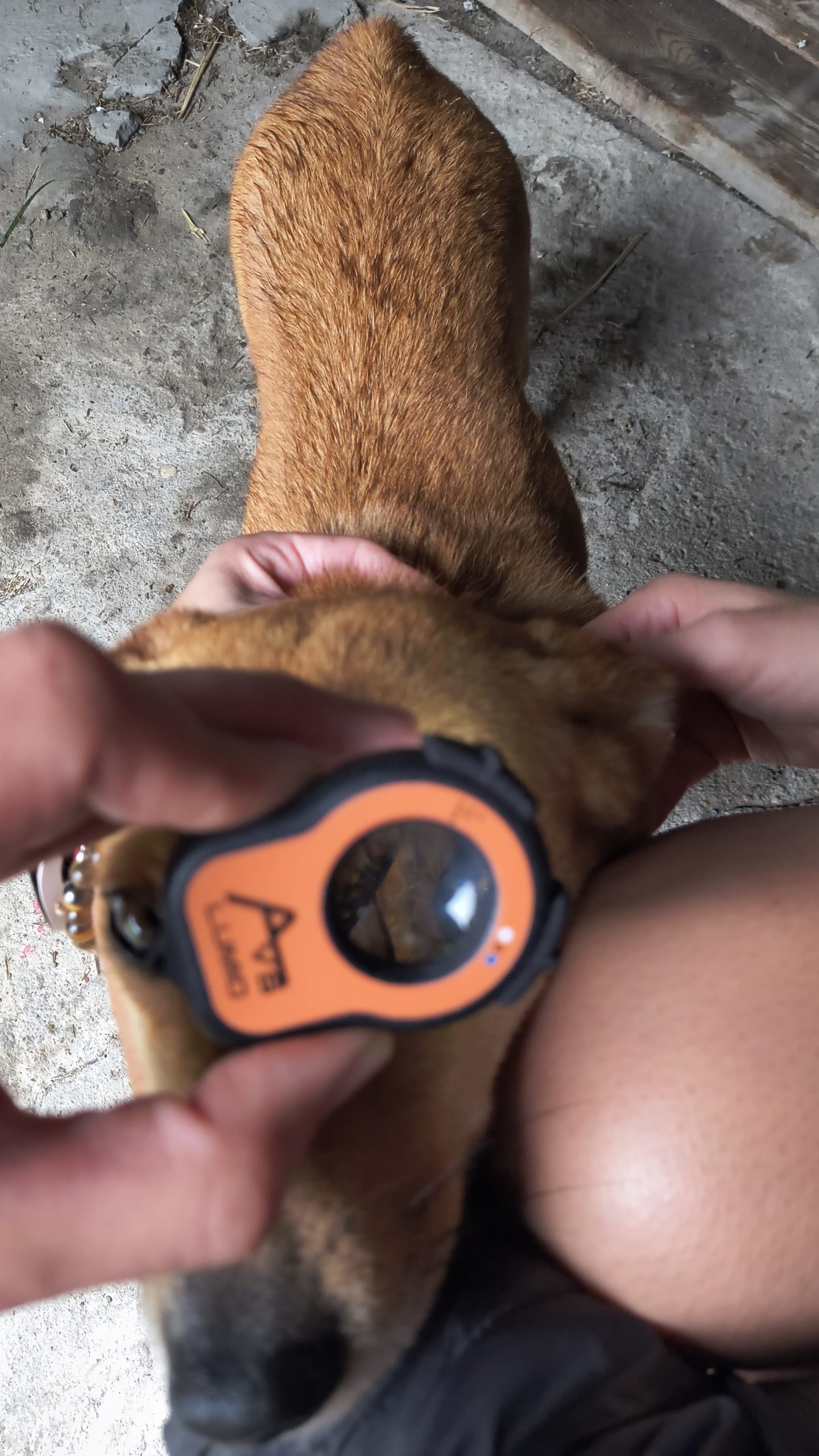
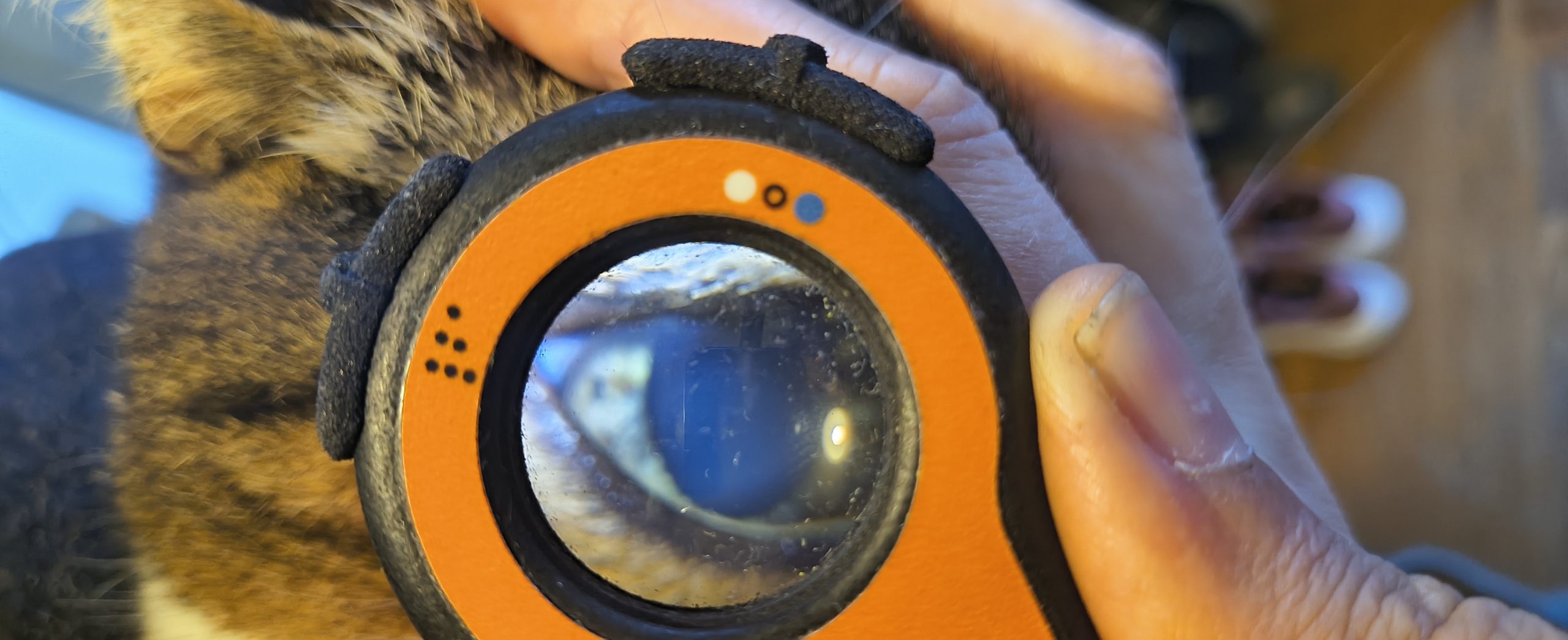
A veterinarian performs a detailed eye examination on a dog using the Aston Vision Sciences Lumio device.
A close-up view of the Lumio device in action during a veterinary eye exam on a dog
A veterinarian positions the Lumio device near a dog’s eye to perform a thorough ophthalmic evaluation
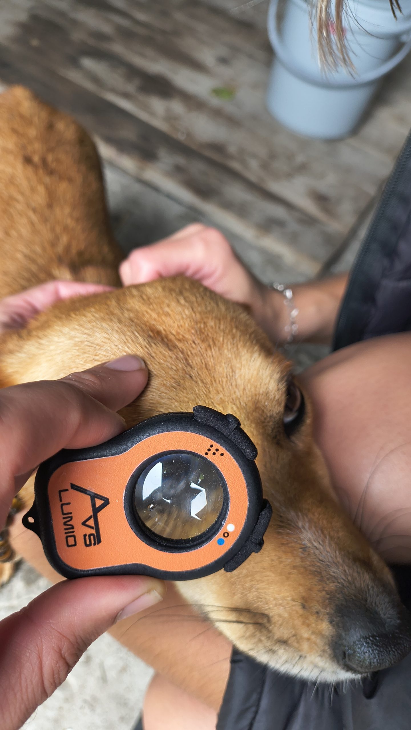
A close-up view of the AVS Lumio device in action, highlighting its use in detailed veterinary eye assessments
A close-up view of the AVS Lumio device in action, highlighting its use in detailed veterinary ocular exams
AVS Lumio device is applied directly to a dog’s eye, enabling detailed visualization of ocular structures during a veterinary assessment.
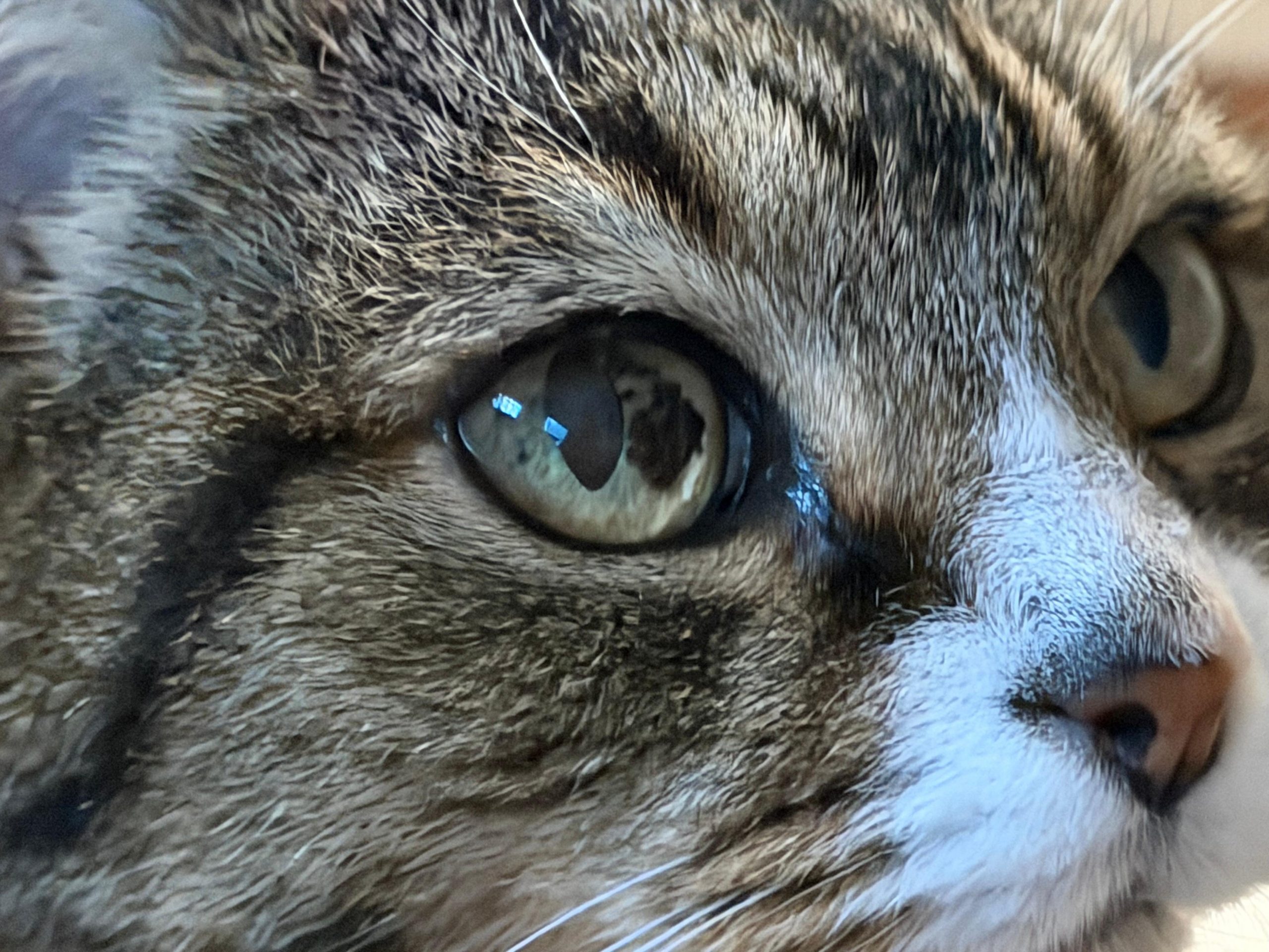
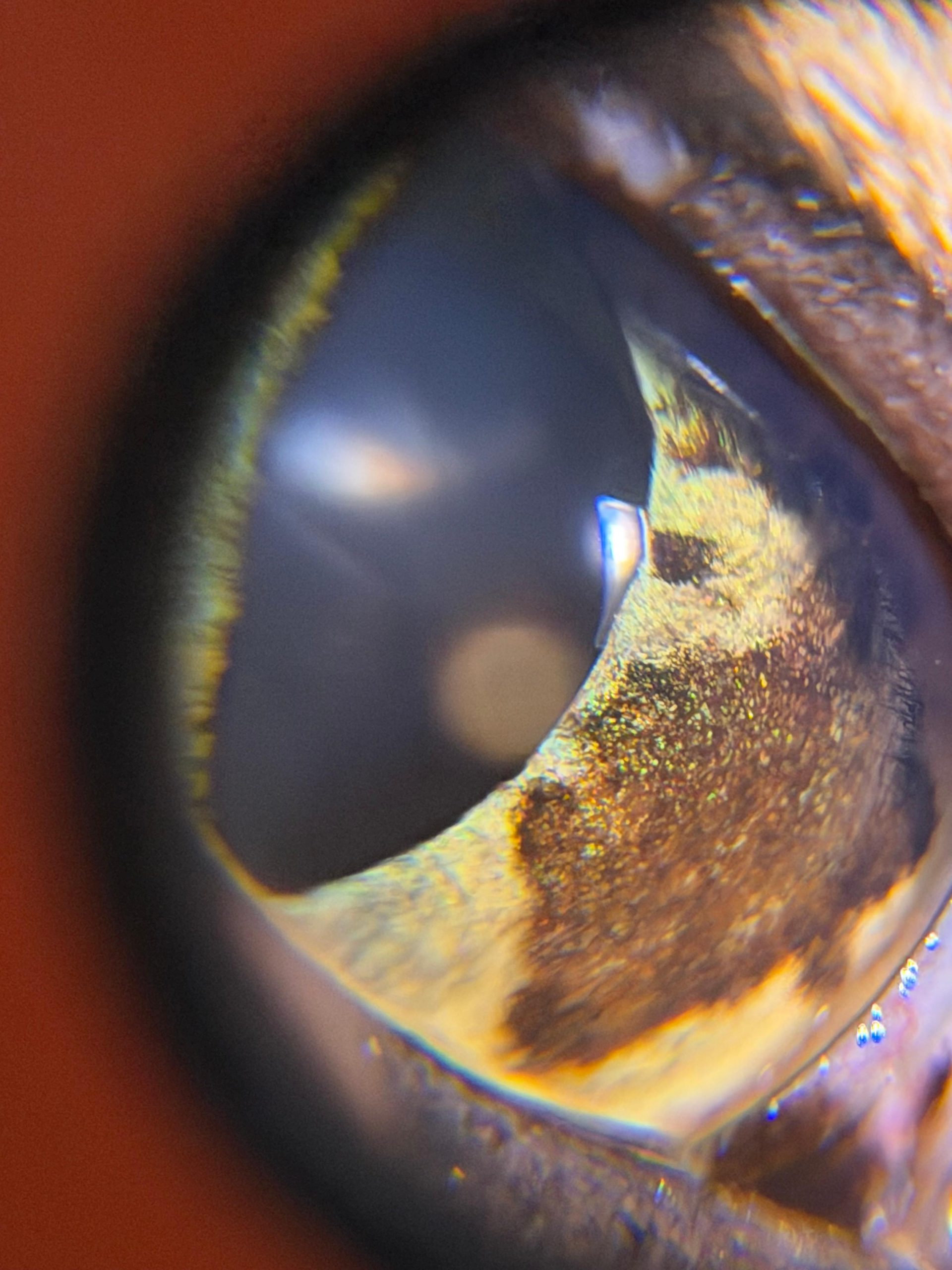
A striking close-up of a tabby cat’s eye, showcasing the beauty of feline iris patterns with unusual but benign Iris Melanosis
Tabby cat anterior eye examination
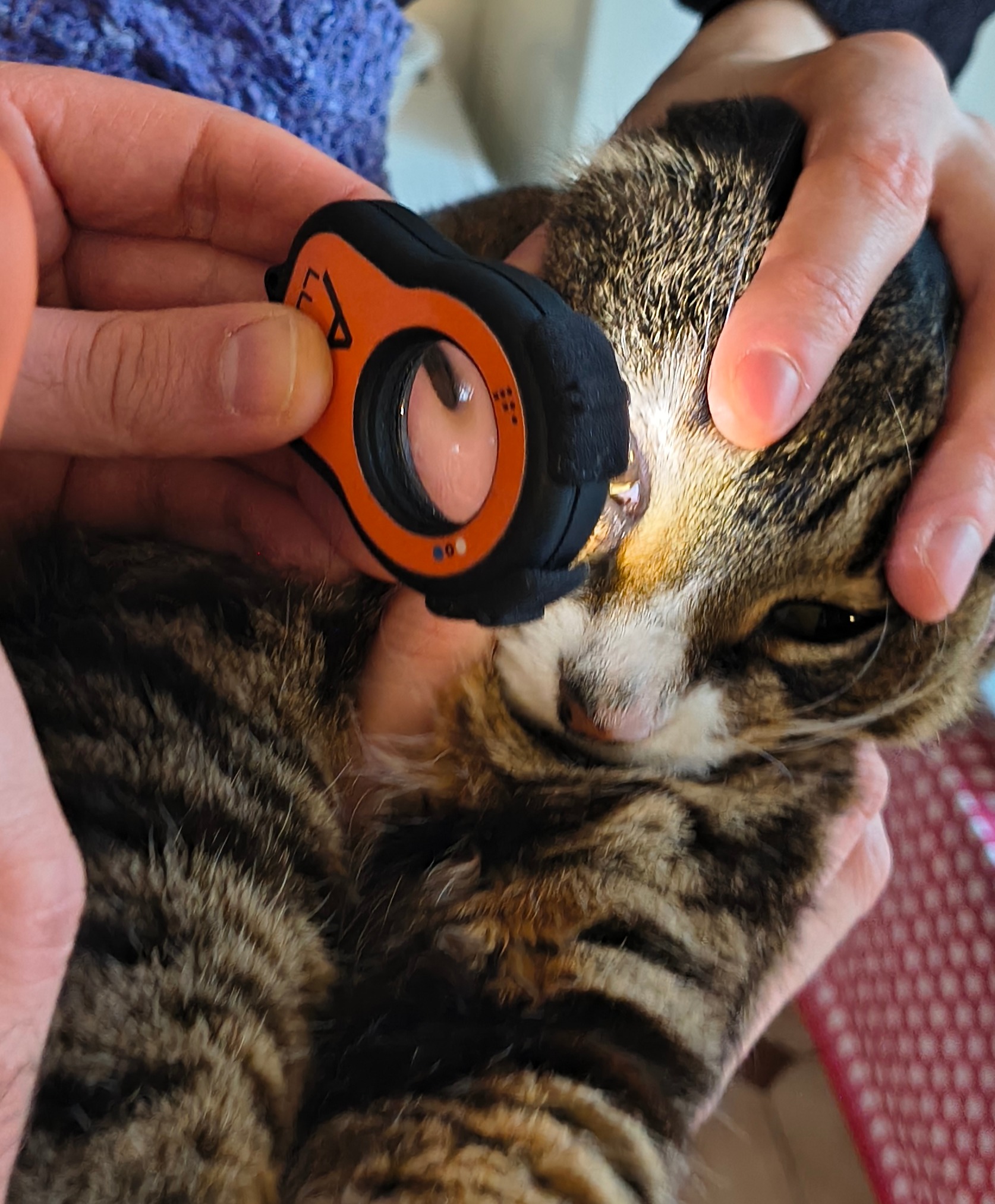
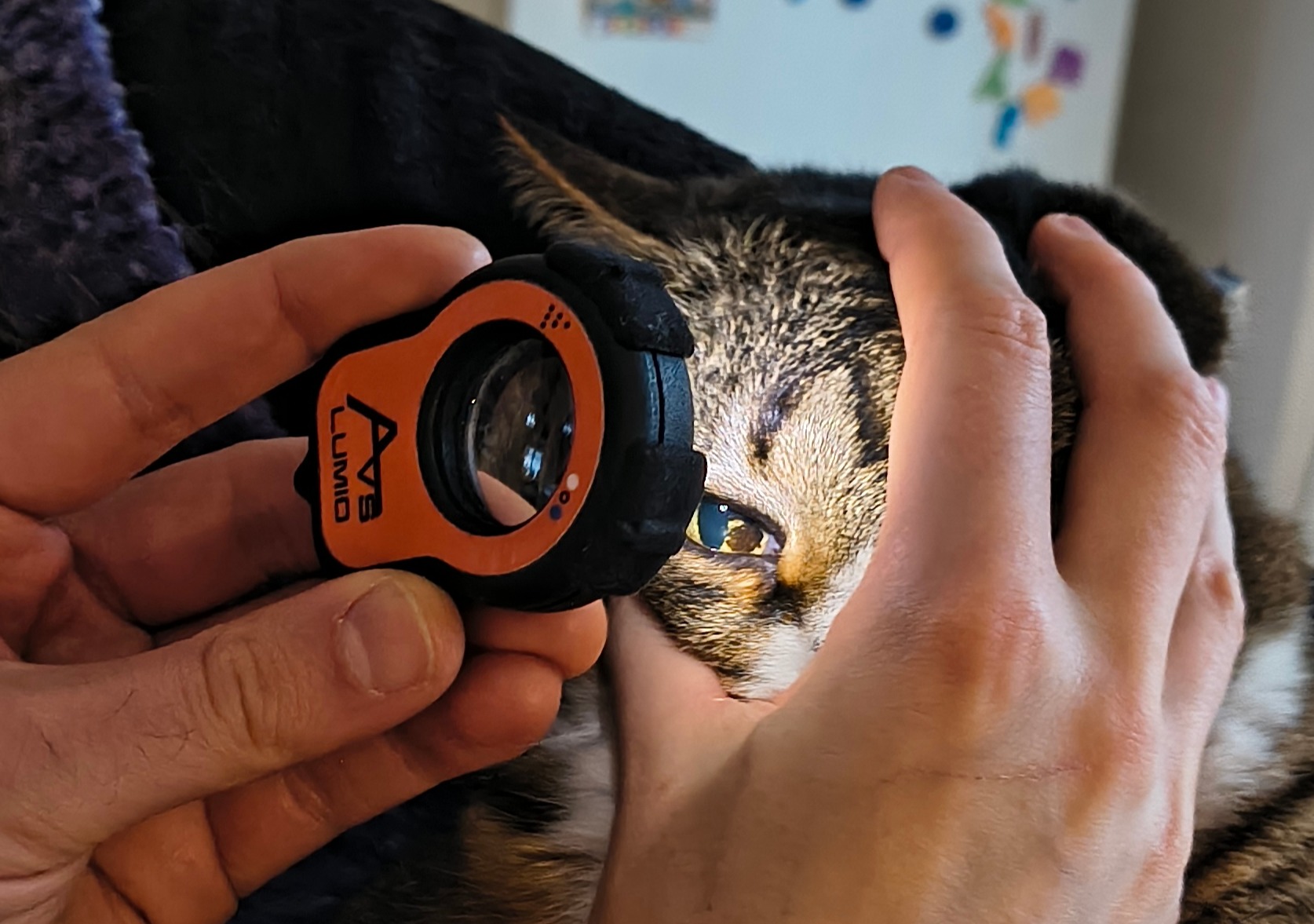
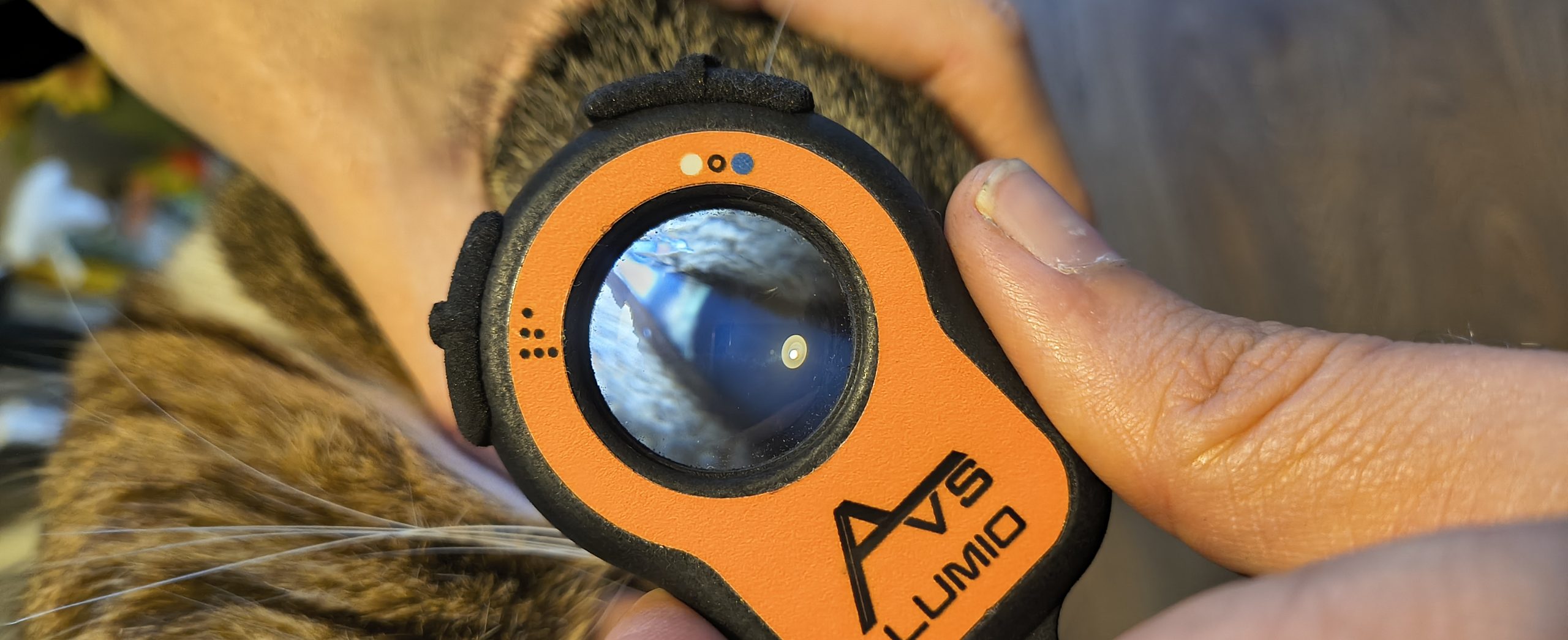
A veterinary professional uses the Lumio device to inspect a cat’s eye, capturing fine structural details.
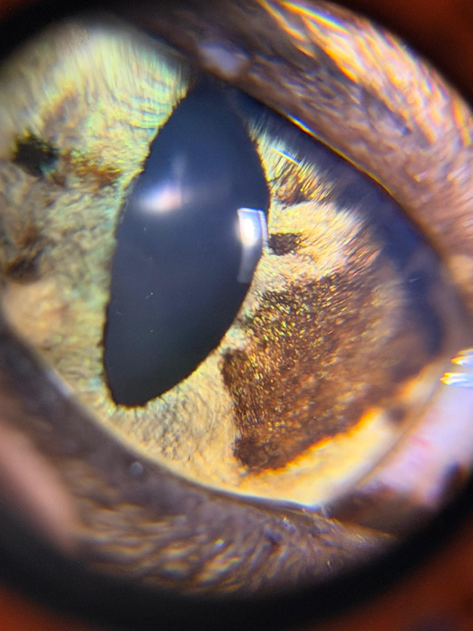
The Lumio device reveals distinct pigmentation on the iris, consistent with iris melanosis, during a veterinary eye examination.
A close-up of a cat’s face during an eye exam using the Aston Vision Sciences Lumio device
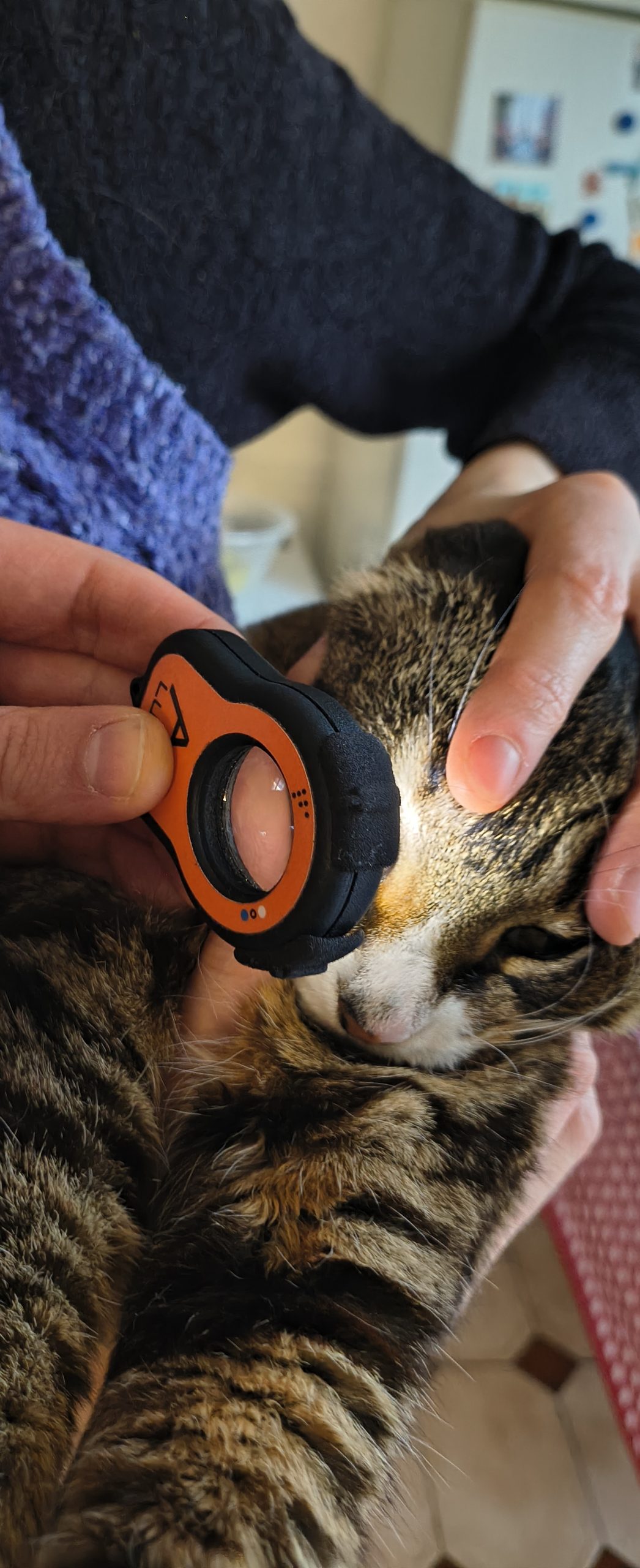
Close-up of a cat’s eye being examined with the Lumio device, highlighting its utility in veterinary ophthalmology
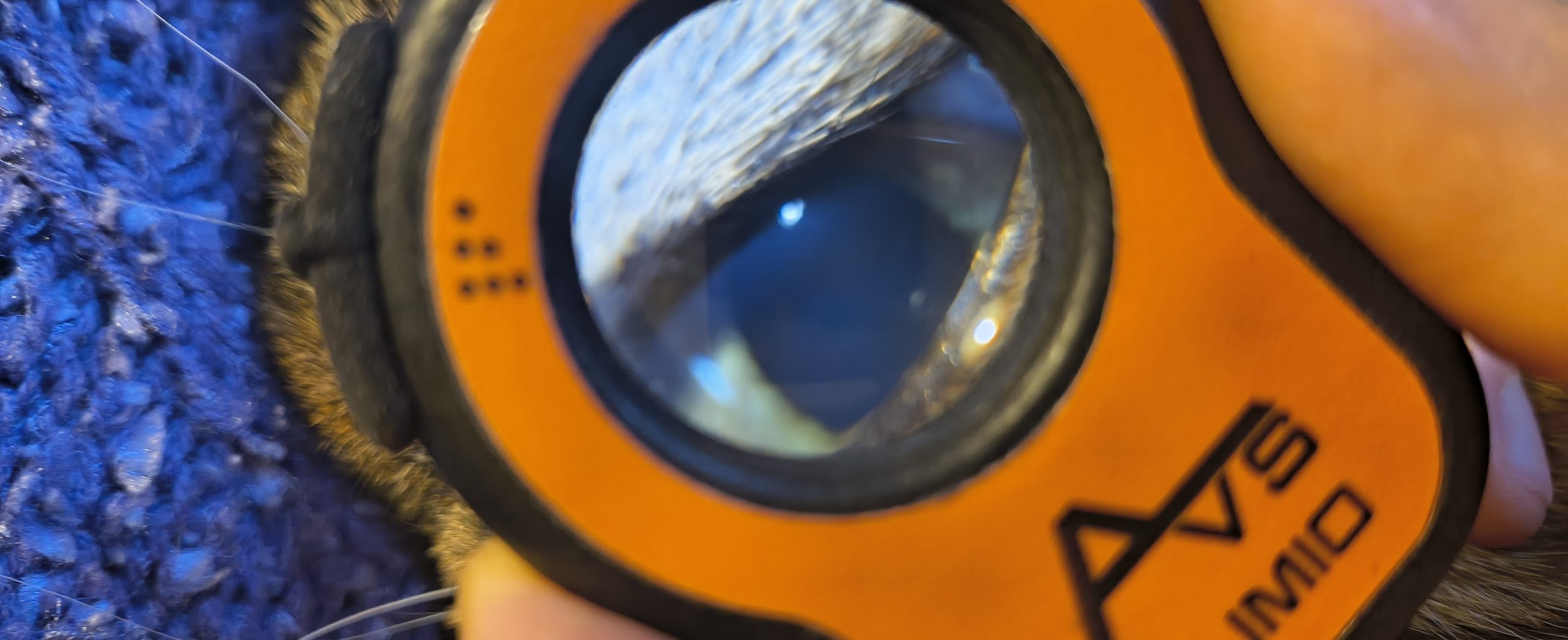
A detailed inspection of a cat’s eye through the Lumio device, revealing ocular surface features for veterinary assessment
The Lumio device is used to perform a focused examination of a cat’s eye, supporting veterinary diagnostics.
The AVS Lumio device is used to examine a cat’s eye under white light, providing a clear view of the crystalline lens for veterinary assessment.
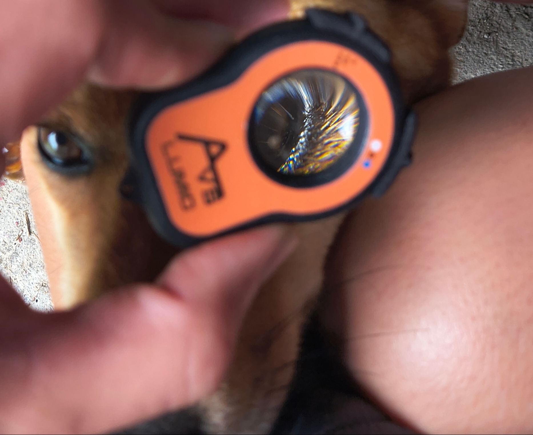
The Lumio device is used to examine a dog’s eye treated with fluorescein dye, revealing corneal surface details under blue light coupled with the onboard fluorescein filter
The Lumio diagnostic tool is used to examine a dog’s eye treated with fluorescein, revealing surface irregularities under blue light illumination.
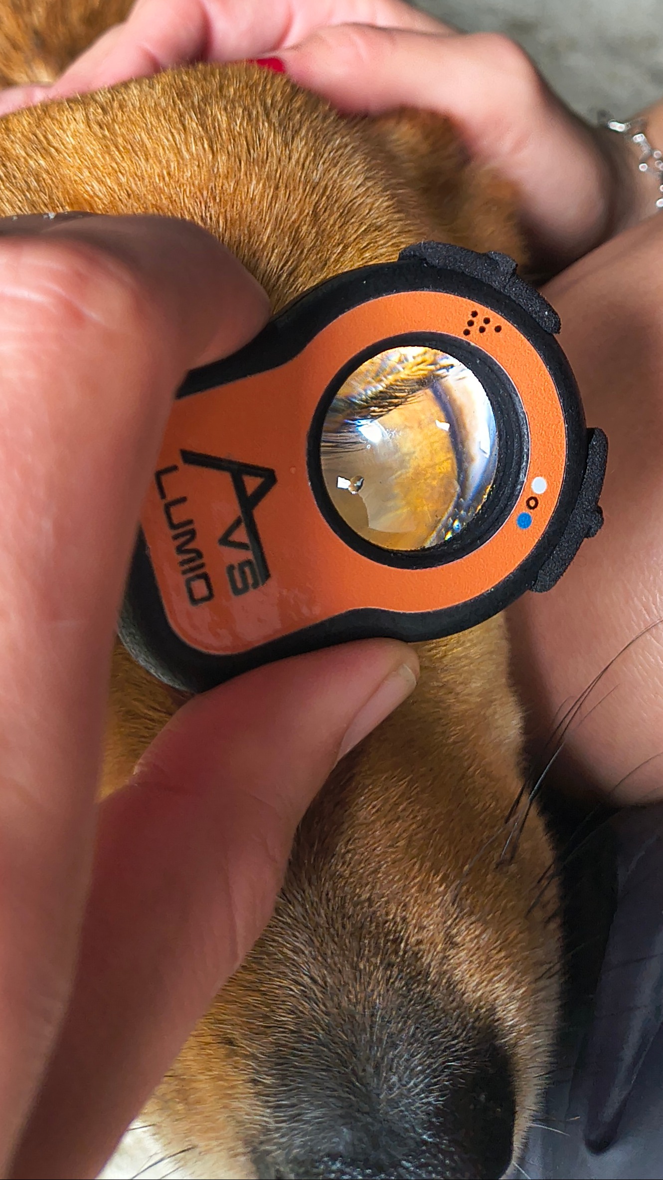
A veterinarian uses the Lumio device to assess a dog’s eye with fluorescein dye, highlighting corneal health in a clinical setting.
A veterinarian uses the Lumio diagnostic device to assess a dog’s eye under fluorescein lighting, helping detect corneal injuries or infections.
A veterinarian uses the Lumio diagnostic device to assess a corneal ulcer present on a dog’s eye under fluorescein lighting.