Revealing the Invisible
Fluorescein is one of the most commonly used dyes in optometry and a cornerstone in ocular diagnostics. Readily obtainable, Fluorescein instillation on to the anterior eye enhances visibility of the tear film, corneal surface, and conjunctiva, allowing clinicians to detect subtle abnormalities that are otherwise invisible in normal white light.
Once introduced on to the ocular surface, Fluorescein requires a wide excitation wavelength of between 420- 520 nm to induce a resultant green fluorescence in the 485- 580 nm region of the colour spectrum allowing for simultaneous corneal and conjunctival qualitative assessment.
- Blue Light Source – Fluorescein fluoresces when excited by blue light in the 465–490 nm range. Unfortunately, many contemporary slit lamps are equipped with sub- optimal blue excitation filters which although are generally within the working spectrum of fluorescein, do not produce maximum viewing performance.
Yellow Barrier Filter – A yellow filter placed in the viewing path, blocks the reflected blue light from the eye and enhances the visibility of the green fluorescence emitted by fluorescein, dramatically improving contrast and detail, making subtle staining patterns easier to detect and interpret.
Modern high- end slit lamp instruments may come furnished with both blue excitation and yellow emission filters built into the body of the devices but viewing performance depends on the quality of the filters and varies considerably from one manufacturer to another. Additionally, many clinics may not have access to quality equipment and rely on alternative low-cost handheld thin film filters which must be manually held in position by the administering clinician during staining tests. This is not particularly comfortable for the clinician and the filters are generally not well tuned to optimum fluorescence emission wavelengths leading to poor viewing experiences and an increased possibility of missed diagnosis.
Why Fluorescein Matters in Eye Care & Dry Eye Disease
Typically, fluorescein allows trained clinicians to identify a whole host of abnormal manifestations such as corneal abrasions, scratches (length and position), corneal corrosions and abscesses, Although considered mildly invasive and has been shown to promote premature breakup in moderate to severe dry eye patients, Fluorescein Breakup Time is one of the most commonly used tests of tear film stability [1, 2, 3]. Lid Wiper Epitheliopathy – thought to be one of the best indicators for the presence of dry eye [4] can also be performed using fluorescein.
Although conjunctival and corneal staining patterns have proved to be particularly effective markers of dry eye disease presence, this is only accurate in the most severe case examples, with less variability reported in both mild to moderate instances of dry eye as compared to healthy control patient [5]. Nevertheless, staining is still seen as one of the preferred techniques by clinicians due to low cost and ease of execution [2, 6].
Enhanced AVS Colour Formula
Traditional fluorescein filters often compromise diagnostic accuracy due to suboptimal spectral transmission — either allowing excessive blue colour to pass through or inadequately isolating the green fluorescence light.
Building on scienced based research conducted during Dr Karl Obszanski’s PhD at Aston University, and further refined at Aston Vision Sciences, we developed a proprietary colour formula designed to isolate Fluorescein emission wavelengths while blocking blue light which may compromise viewing. By refining the spectral cut-off characteristics and maximizing transmissivity, our filter enhances contrast and suppresses background glare, enabling clearer visualization of tear film instability, epithelial staining, and subtle anterior segment changes, supporting more confident clinical decision-making, particularly in dry eye diagnostics and corneal surface evaluation.
Improved Diagnostic Accuracy
Clearer detection of tear film instability, epithelial defects, and subtle anterior segment changes — critical in dry eye and corneal assessments
Optimised Spectral Transmission
Enables clearer detection of tear film instability, epithelial defects, and subtle anterior segment changes — critical in dry eye and corneal assessments
Enhanced Contrast and Clarity
Engineered to Fluorescein emission wavelengths, enhancing green colour visibility, even at lower blue light brightness levels
Reduced Visual Noise
Suppress excess blue light which obscures fine details allowing for greater confidence and decision making when examing
Filter Comparison
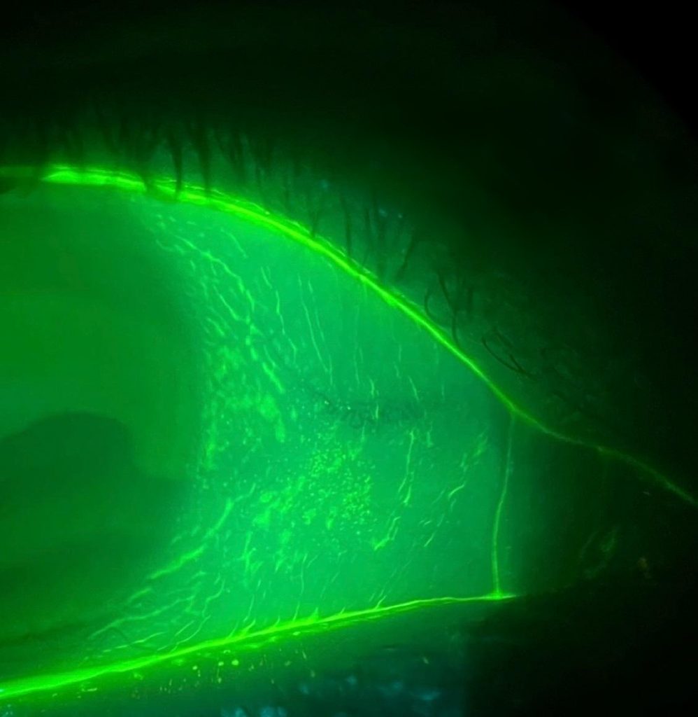
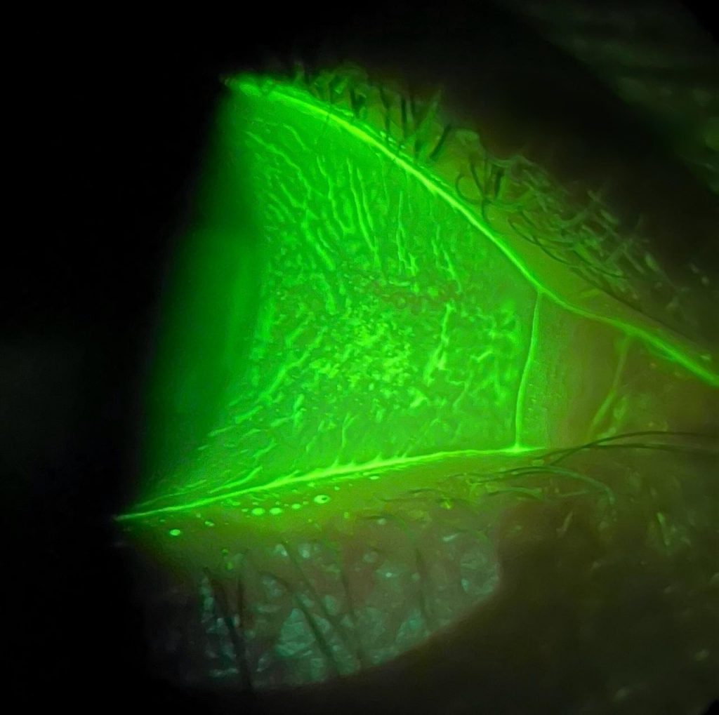
Independent images submitted by customer on the same patient, images taken one after another in close time proximity

Regular Filter
A view of the anterior segment of the eye captured by using a non- optimised fluorescein filter. Details are reduced, blurry and colour tone shows a blue tinge suggesting blue light leakage through a less than optimal filter.

AVS Optimised Filter
View of Conjuntival Folds and Episcleral Vessels become more pronounced when viewed with AVS filter which enhances contrast, making Superficial blood vessels, Conjunctival microfolds, Surface irregularities more visible.
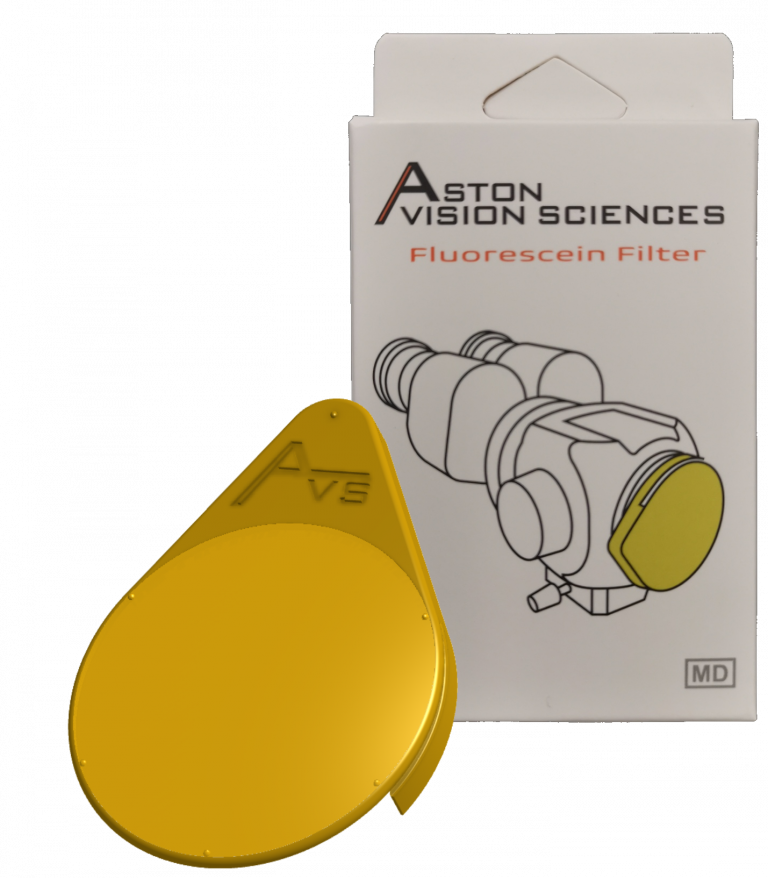
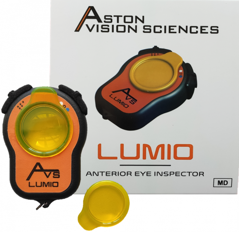
By merging innovation with ophthalmic needs, AVS developed two cutting edge products designed with clinical and patient needs in mind: the clip on Fluorescein Filter, the portable LumioAnterior Eye Examiner, with more clinically drivern devices coming.
Interested In Industrial Use?
Our colour formula can be used in other Fluorescein related applications such as leak detection, flow analysis, forensic and environmental.
Contact us if you want to explore more.
References
- M.S. Norn, Desiccation of the Precorneal Film, Acta Ophthalmologica 47(4) (1969) 865-880.
- A.W. Turner, C.J. Layton, A.J. Bron, Survey of eye practitioners’ attitudes towards diagnostic tests and therapies for dry eye disease, Clin. Exp. Ophthalmol. 33(4) (2005) 351-355.
- L.S. Mengher, A.J. Bron, S.R. Tonge, D.J. Gilbert, Effect of fluorescein instillation on the precorneal tear film stability, Curr. Eye Res. 4(1) (1985) 9-12.
- D.R. Korb, J.P. Herman, C.A. Blackie, R.C. Scaffidi, J.V. Greiner, J.M. Exford, V.M. Finnemore, Prevalence of Lid Wiper Epitheliopathy in Subjects With Dry Eye Signs and Symptoms, Investigative Ophthalmology & Visual Science 50(13) (2009) 4640-4640.
- J.S. Wolffsohn, R. Arita, R. Chalmers, A. Djalilian, M. Dogru, K. Dumbleton, P.K. Gupta, P. Karpecki, S. Lazreg, H. Pult, B.D. Sullivan, A. Tomlinson, L. Tong, E. Villani, K.C. Yoon, L. Jones, J.P. Craig, TFOS DEWS II Diagnostic Methodology report, Ocular Surface 15(3) (2017) 539-574.
- D.R. Korb, Survey of preferred tests for diagnosis of the tear film and dry eye, Cornea 19 (4) (2000) 483-486


